
Abbreviations
LMS: Leiomyosarcoma
α-SMA: α-Smooth Muscle Actin
Introduction
Soft tissue sarcomas in dogs are characterized as mesenchymal tumors with local invasion, encompassing a diverse array of mesenchymal tumors exhibiting different histogenetic origins, and they constitute approximately 15% of all dermal and subdermal neoplasms in canines. Typically, mature or older dogs demonstrate a higher prevalence, with larger breeds showing an increased vulnerability. Anatomically, nearly 60% of soft tissue sarcomas in dogs are located on motor organs. In contrast, other somatic regions exhibit a comparatively lower incidence and for example the torso (35%) and head and neck (5%) are less frequently affected [ 1 ]. Malignant cutaneous smooth muscle tumors are uncommon in both humans and animals. These tumors possibly arise from the smooth muscles that erect the hair, those inside the vascular septum, or from special muscles in the genital region [ 2 ]. Cutaneous leiomyosarcoma is uncommon in both humans and animals. While multiple well-differentiated smooth muscle tumors have been reported in dogs, cats, ferrets, and horses, cutaneous smooth muscle neoplasms are typically solitary in nature [ 3 ]. Histologically, LMS is characterized by intertwined neoplastic smooth muscle cells. In humans, malignant smooth muscle tumors display marked proliferative activity, cellular uniformity, nuclear atypia, and intratumor necrosis. Human LMS is further classified into myxoid, epithelioid, and pleomorphic subtypes, while the last-mentioned subtype was only diagnosed in animal health publications, as some of them were similar to anaplastic sarcomas [ 2 ]. In human medicine, leiomyosarcoma is associated with high rates of recurrence and metastasis, resulting in a generally poor prognosis. The most frequent metastatic sites include the lungs, bone, brain, and lymph nodes. Surgical removal is the most effective treatment for leiomyosarcoma, while chemotherapy does not appear to be as effective, is largely palliative, and is typically reserved for patients with extensive metastasis. Previous reports revealed an acceptable prognosis for surgically treated dogs with spleen, stomach, small intestine, and especially the cecum leiomyosarcomas. Nevertheless, the number of studies on dogs remains limited. The anatomical site of leiomyosarcoma in dogs holds greater prognostic significance compared to human. Leiomyosarcomas originating from superficial cutaneous locations are associated with a more favorable prognosis. In contrast, osseous involvement of the human leiomyosarcoma represents a potential negative prognostic indicator.[ 4 ]. β-Catenin, encoded by the ctnnb1 gene, is a key component of intercellular junctions, and dysregulation of its expression has been implicated in the progression of animal and human tumors. Studies have demonstrated that disruption of the E-cadherin/β-catenin complex regulations affects both types of canine melanotic tumors, also disruption of the E-cadherin/β-catenin complex and increased β-catenin levels can lead to tumor progression and malignancy [ 5 ]. In conventional mature cutaneous cells, β-catenin constitutes a fundamental component of the intercellular junctions, while the Wnt/β-catenin signalling cascade significantly influences the maintenance of skin homeostasis, particularly in the preservation of hair follicle stem cells. However, data regarding the expression profile of β-catenin in normal dermal tissue, as well as in epidermal and follicular neoplasms in canines remain scarce. In malignant neoplasms, including squamous cell carcinoma, basal cell carcinoma, sebaceous gland carcinoma, apocrine gland carcinoma, and epithelial carcinoma, reduced or absent membranous β-catenin expression has been reported, suggesting that the decrease or loss of β-catenin expression is pivotal in the development of a malignant phenotype and may play a role in the invasive behaviour or metastasis of these carcinomas [ 6 ]. Cadherins are multi-family membrane glycoproteins responsible for calcium-mediated homophilic attachment. They are particularly related to the joint area and play roles in processes including tissue morphogenesis and tumorigenesis in complex organisms. E-cadherin, the predominant cadherin expressed in epithelial tissues, is widely regarded as an anti-oncogene and a developmental agent in epithelium-originated neoplasms. E-cadherin downregulation has been associated with decreased cellular differentiation, enhanced invasiveness , and metastatic behavior in a variety malignancies, including several sarcomas. Epidermal growth factor receptor (EGFR), commonly referred to as HER1 or erbB1, constitutes a member of the receptor tyrosine kinase family. Besides other ErbB family members, including ErbB2, ErbB3, and ErbB4, they can form heterodimers. These receptors are integral to essential cellular functions, encompassing cell division and specialization. Overexpression of COX-2 and EGFR has been documented in numerous malignant neoplasms. These molecules contributes to critical processes at several pivotal stages, including vascularization, inhibition of programmed cell death, suppression of immune responses, enhancement of cell division, possibility for progression, cellular specialization, and displacement. COX-2 and EGFR represent promising targets for therapeutic and chemotherapeutic interventions in the treatment of diverse pathological conditions, such as neoplasm. Therefore, due to their involvement in tumor invasiveness, malignancy, reduced survival rates, and poor prognostic, they are considered promising biomarkers and potential therapeutic targets in veterinary oncology [ 7 ]. The present study aimed to investigated the differential diagnosis of cutaneous leiomyosarcoma tumors in dogs, and the changes in the immunoexpressing of adhesion markers, including E-Cadherin, β-Catenin, in addition to the changes in the expression of markers CK, MyoD1, Myogenin, CD31, Desmin, α-SMA, Vimentin, Ki67, and EGFR in the local and invasive forms of these tumors.
Results
Skin lesions
A total of 318 dogs presenting with various diseases and disorders were examined. These included parasitic lesions (scabies) (n = 49/318, 15.40%), fungal infection (dermatophytosis) (n = 42/318, 15.20%), traumatic lesions (skin tear or wound) (n = 13/318, 4.08%), papilloma (n = 27/318, 8.49%), fibroma (n = 8/318, 2.51%), fibrosarcoma (n = 6/318, 1.88%), leiomyosarcoma (n = 6/318, 1.88%), trichoblastoma (n = 3/318, 0.94%), and squamous cell carcinoma (SCC) (n = 2/318, 0.62%). The remaining cases (n = 162/318, 50.94%) were associated with various clinical manifestations of systemic diseases and/or allergic dermatopathies.
Pathological Findings
Among the examined dogs, 6 cases were diagnosed as leiomyosarcoma (LMS) tumors, including 3 cases of localized cutaneous leiomyosarcoma (local LMS), and 3 cases of invasive cutaneous leiomyosarcoma (invasive LMS). In the local LMS group, affected dogs included one male Rottweiler, one female Terrier, and one female Iranian mixed breed dog. Their ages ranged from 6 to 10 years, and tumor diameters varied between 3 and 37 mm. Tumor locations were mainly in the lower limbs (locomotor), neck, and abdomen. In the invasive LMS group, one male Iraqi breed dog, one female Shih Tzu, and one male German Shepherd were affected. These dogs were aged between 6 and 12 years, with tumor sizes ranging from 2 to 26 mm. Tumors were primarily located in lower limbs (locomotor), neck, and abdomen regions. Metastasis spread to the conjunctiva, pulmonary system, or gastrointestinal tract was confirmed through histopathological examination (Figure 1). Histological evaluation of H&E and Masson's trichrome stained sections revealed a moderate degree of cellular and nuclear pleomorphism in both local and invasive LMS, with the nucleoli count in tumor cells ranging from 2 to 4, and the mean mitotic index for local and invasive LMS was 4.2 and 5.8, respectively. Neoplastic smooth muscle fibres exhibited an interwoven configuration characterized by an irregular morphology, with minimal intervening connective tissue. Necrosis or hemorrhage were absent in local LMS, but were observed in invasive LMS (Figure 2).
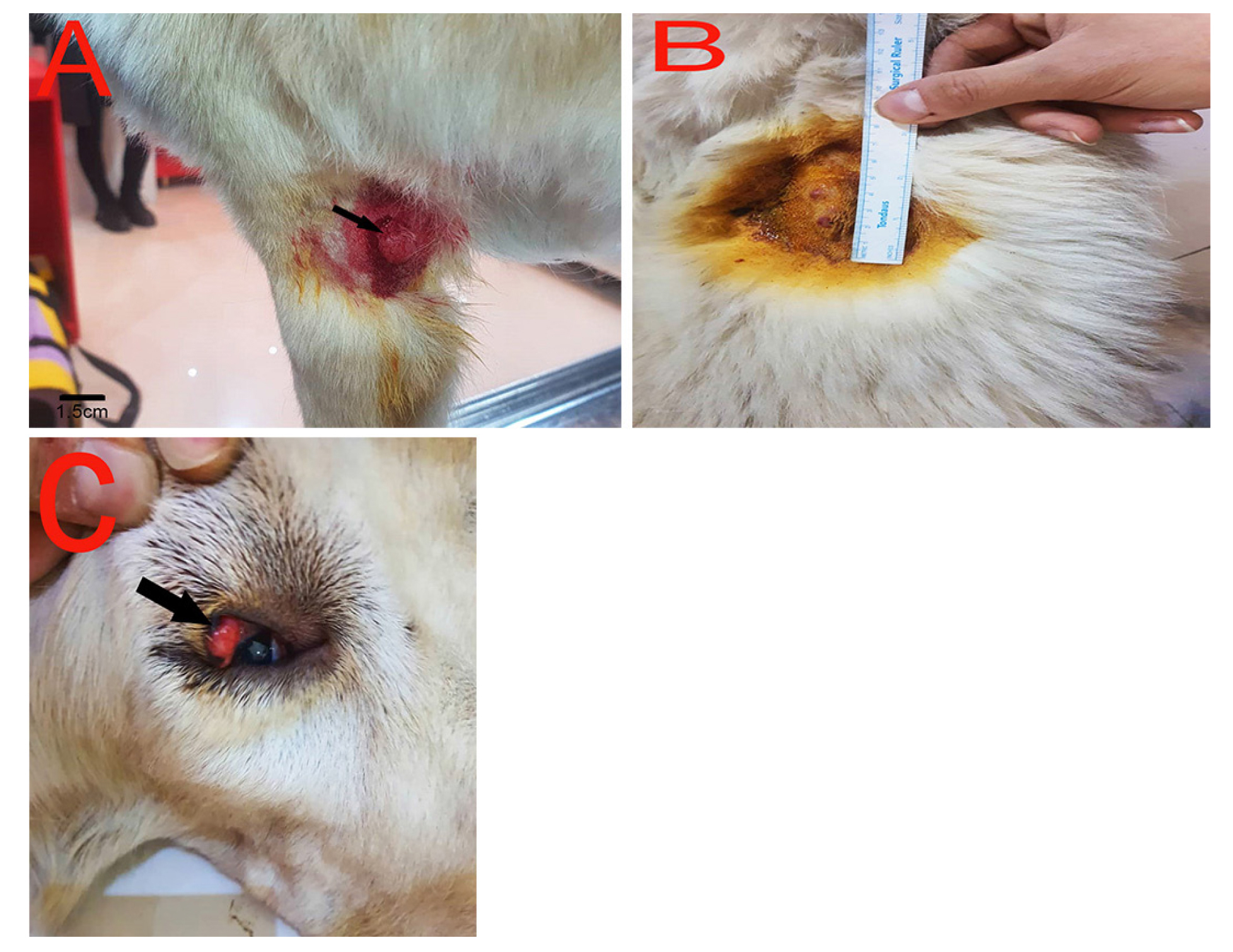
Figure 1. The appearance of the local and invasive LMS tumor in dogs. A: A local LMS tumor, 1.5 cm in diameter, of pink color, and a limited formation in the skin of the foot of an 8-year-old female Iranian mixed-breed dog. B: Invasive LMS with pink nodular masses with 2 to 7 mm diameters in various parts of the skin of a 6-year-old male Iraqi breed dog. C: Presence of invasive LMS tumor mass inside the conjunctiva of the Iraqi dog breed, with a diameter of about 1 cm.
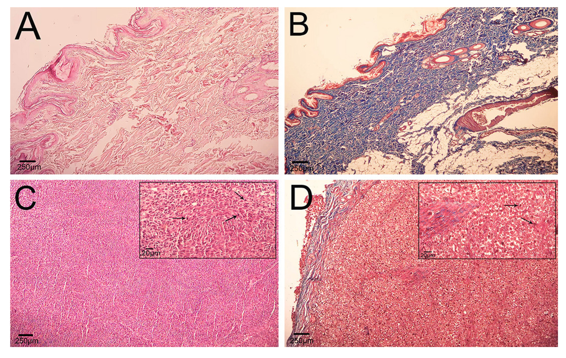
Figure 2.Light microscope photomicrographs of LMS tumors compared with normal tissue of dog skin.A: Organized structure of epidermis and dermis of normal dog skin (H&E). B: Organized structure of normal dog skin, despite the connective tissue (blue), dermis, and smooth muscles (red) around the blood vessels (Masson's trichrome). C: Cutaneous LMS with a view of interwoven fibers of smooth muscle, and the whirlwind view with cells formed and cut in various directions. The small picture presents the smooth muscle tumor cells with cellular pleomorphism, and the presence of mitotic figures (arrow) (H&E). D: A view of a section of LMS tumor where muscular tumor cells are visible (red) with limited connective tissue (blue) inside the tumor parenchyma. The small picture shows various forms of tumor cells and mitotic status (arrow) (Masson's trichrome).
Hematological Findings
One-way ANOVA analysis demonstrated no significant difference in mean values of MCV, PLT, Neutrophils, and Lymphocytes among healthy dogs and those with local or invasive LMS (p > 0.05). In contrast, significant difference were detected in Hct, Hb, RBC, MCHC, WBC, Monocytes, and Eosinophils among healthy dogs and those with invasive LMS (p < 0.05). Within the local LMS group, mean value for RBC, MCHC, WBC, and Eosinophils did not differ significantly compared to healthy dogs (p > 0.05). Detailed hematological data are summarized in Table 1.
| Parameter | Dogs (n= 12) | p value | ||
|---|---|---|---|---|
| Normal (n= 6) | Local LMS (n= 3) | Invasive LMS (n= 3) | ||
| HCT (%) (Reference: 37-55) | 44.80 ± 4.82b | 37.60 ± 3.05 a | 34.20 ± 2.39 a | 0.002 |
| Hb (g/l) (Reference: 140-190) | 161.40 ± 9.24 b | 147.20 ± 11.30 ab | 137.00 ± 8.36 a | 0.006 |
| RBC (1012/l) (Reference: 5.8-8.50) | 6.92 ± 0.87 b | 6.96 ± 0.65 b | 5.66 ± 0.61 a | 0.023 |
| MCV (ƒl) (Reference: 66-75) | 69.20 ± 1.64 | 69.20 ± 1.48 | 70.40 ± 3.78 | 0.695 |
| MCHC (g/l) (Reference: 32-36) | 33.00 ± 1.58 b | 33.40 ± 2.07 b | 30.00 ± 1.58 a | 0.019 |
| PLT (109/l) (Reference: 150-400) | 251.40 ± 58.44 | 240.00 ± 56.51 | 228.60 ± 67.34 | 0.841 |
| WBC (109/l) (Reference: 6-13) | 8.92 ± 1.25 a | 7.84 ± 0.76 a | 13.42 ± 2.49 b | 0.001 |
| Neut (109/l ) (Reference: 3-10.50) | 6.76 ± 1.19 | 6.44 ± 1.23 | 6.24 ± 1.32 | 0.806 |
| Lym (109/l) (Reference:1-4) | 2.36 ± 0.68 | 2.66 ± 0.40 | 2.72 ± 0.75 | 0.637 |
| Mon (109/l) (Reference: 0.15-1.2) | 0.52 ± 0.28 a | 0.62 ± 0.25 ab | 1.18 ± 0.46 b | 0.023 |
| Eos (109/l) (Reference:0-0.1.3) | 0.40 ± 0.20 a | 1.12 ± 0.37 b | 1.34 ± 0.46 b | 0.004 |
| Different superscript letters (a, b, ab) designate a notable difference in every row. p <0.05 is significant. | ||||
Immunohistochemical Findings
Following definitive diagnosis, immunohistochemistry staining for CD31 was negative in all six leiomyosarcoma cases. Hence, it was confirmed that all diagnosed LMS cases originated from the smooth muscles responsible for erecting hair (Figures 3 and 4). Additionally, MyoD1 and Myogenin immunohistochemical was also negative for all tumors samples that the nature of the tumors was diagnosed as definite LMS. One-way ANOVA showed no significant differences in the expression percentage or H-score of MyoD1, Myogenin, and CD 31 between healthy dogs and those with local or invasive LMS (p > 0.05) (Figures 3 and 4).
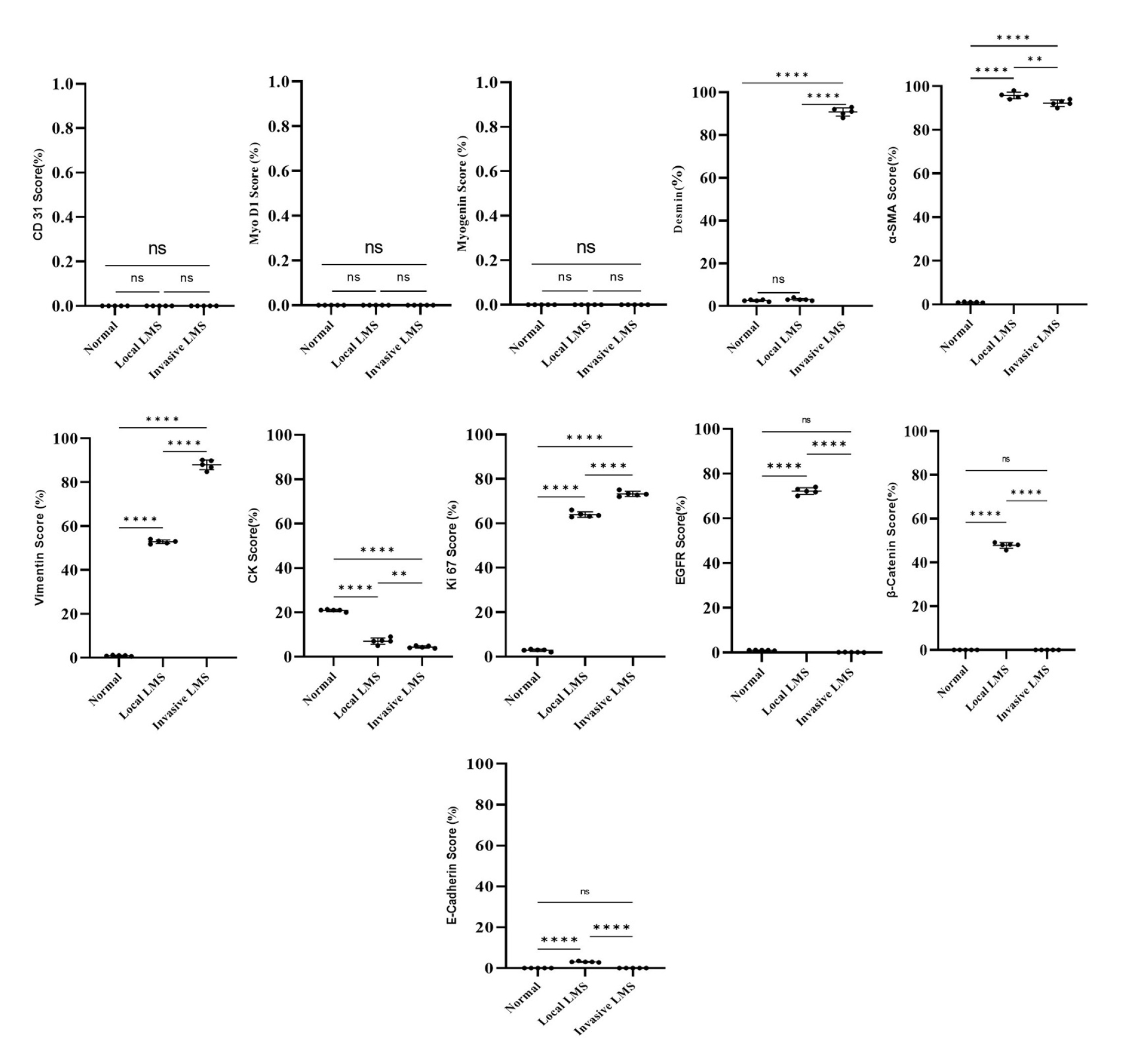
Figure 3.The results of the one-way ANOVA for the percentage of cellular expression of various immunohistochemical markers in the normal, local LMS and invasive LMS groups. Data are presented as Mean ± SD. *: Significant value (p < 0.05). ns: non-significant.
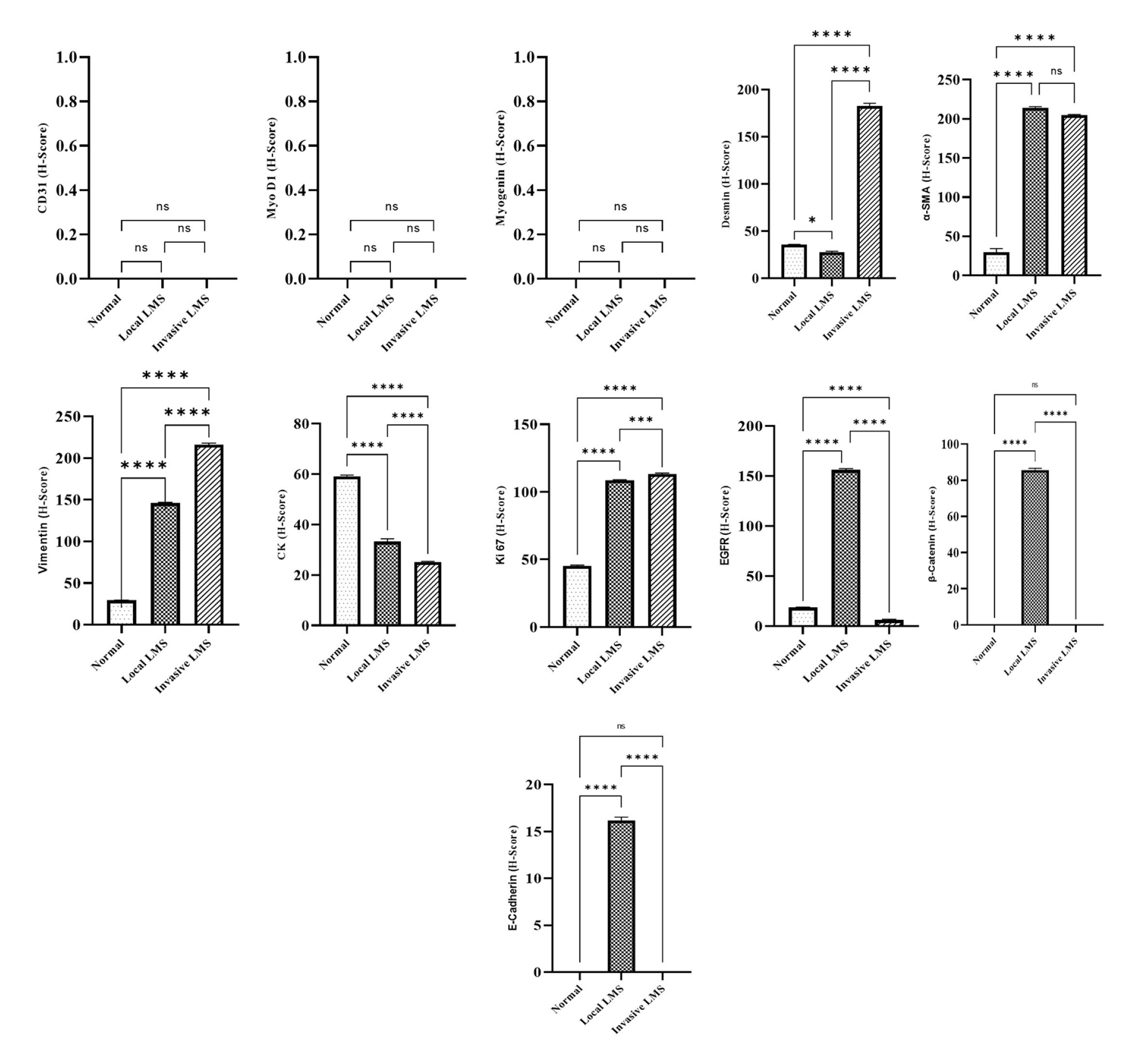
Figure 4.The results of the one-way ANOVA for the H-Score of the different immunohistochemical markers in the normal, local LMS and invasive LMS groups. Data are presented as Mean ± SD. *: Significant value (p < 0.05). ns: non-significant.
In contrast, immunohistochemical analysis showed a significant increase in the percentage of Desmin-positive cells in the invasive LMS group compared to the normal skin, and significant difference were observed in H-Scores among all three groups (p < 0.001) (Figures 3 and 4). The increment of the number of α-SMA+ cells in the LMS groups (local and invasive) compared with the normal animals was significant, and the difference in the H-Scores of the LMS groups (local and invasive) in comparison with the normal category was also significant (p < 0.001). However, no significant difference was observed between local and invasive LMS groups (p > 0.05) (Figures 3 and 4). Vimentin expression was significantly increased, also significant difference was observed in H-Score of the LMS groups compared with the normal animals (p < 0.001) (Figures 3 and 4). Conversely, a significant reduction in the number of CK+ cells and the difference in the H-Score were observed in both LMS group compared with the normal animals (p < 0.001) (Figures 3 and 4). The number of Ki67-positive cells and the difference in the H-Score of the LMS groups compared with the normal animals were significantly increased (p < 0.001) (Figures 3 and 4). The number of EGFR-positive cells in the local LMS group compared with the normal and invasive LMS group was significantly Increased; also, the difference in the H-Score of the three groups was significant (p < 0.001). Notably, EGFR expression percentage and H-Score were significantly reduced in invasive LMS group compared with the other two groups (Figures 3 and 4). β-Catenin expression followed a pattern similar to that of EGFR, with significantly higher expression in local LMS compared with normal and invasive LMS, and significant difference in H-Score among all groups (p < 0.001) (Figures 3 and 4). E-Cadherin expression demonstrated a similarity with β-Catenin and EGFR, although the expression percentage and H-Score in the local LMS group were lower than those observed for EGFR and β-Catenin (Figures 3 and 4). Immunohistochemical expression patterns of all evaluated markers in the normal, local LMS and Invasive LMS are presented in Figures 5- 7.
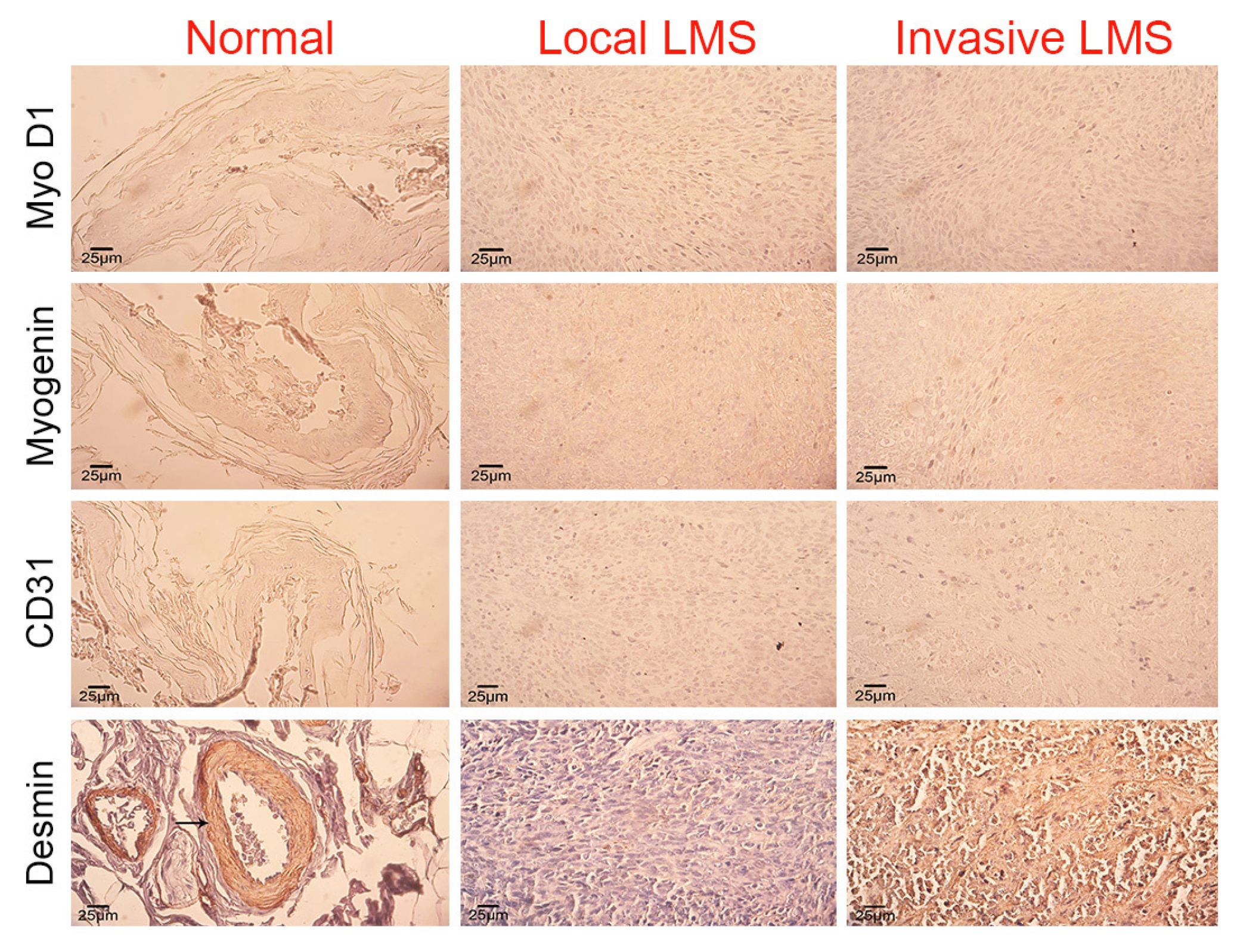
Figure 5.Comparative immunohistochemical photomicrographs of normal skin tissue, local LMS, and invasive LMS. The lack of MyoD1 and Myogenin markers in the normal and tumor tissue cells confirmed the LMS tumors and differentiated them from rhabdomyosarcoma tumors. Lack of CD31 marker expression in LMS tumors indicates the confirmation of piloleiomyosarcoma tumors. Desmin can be seen in normal tissue and in muscles surrounding the blood vessels (the arrow), and while it is not expressed in the local LMS, it can be seen in a dispersed form in invasive LMS (IHC).
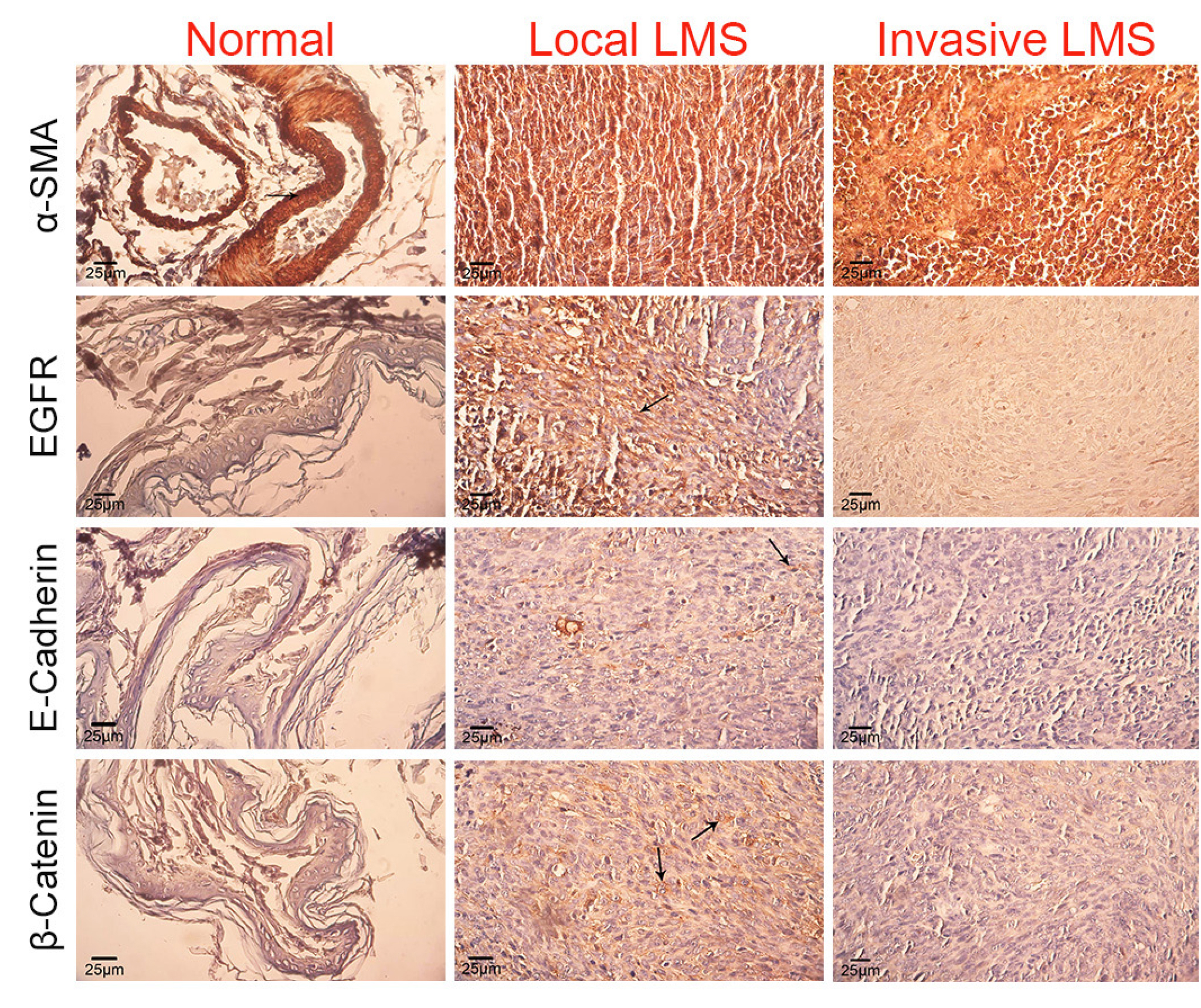
Figure 6.The following comparative immunohistochemical photomicrographs of normal skin tissue, local and invasive LMS.The strong and dispersed α-SMA expression in the local and invasive tumor is an indication of their smooth muscle origin for the tumor-forming cells, and its expression in normal skin can be observed only in smooth muscles around the blood vessels. EGFR expression in normal cells is negative; yet, cytoplasmic expression of EGFR (arrow) can be seen, while the marker is not expressed in the invasive tumor. E-Cadherin expression in normal skin and invasive tumor is negative, but a weak cytoplasmic expression (arrow) can be seen in the local tumor. β-catenin expression is negative in normal skin and invasive tumor; however, a cytoplasmic expression (arrow) of β-catenin in the local tumor can be seen, which is higher than E-Cadherin (IHC).
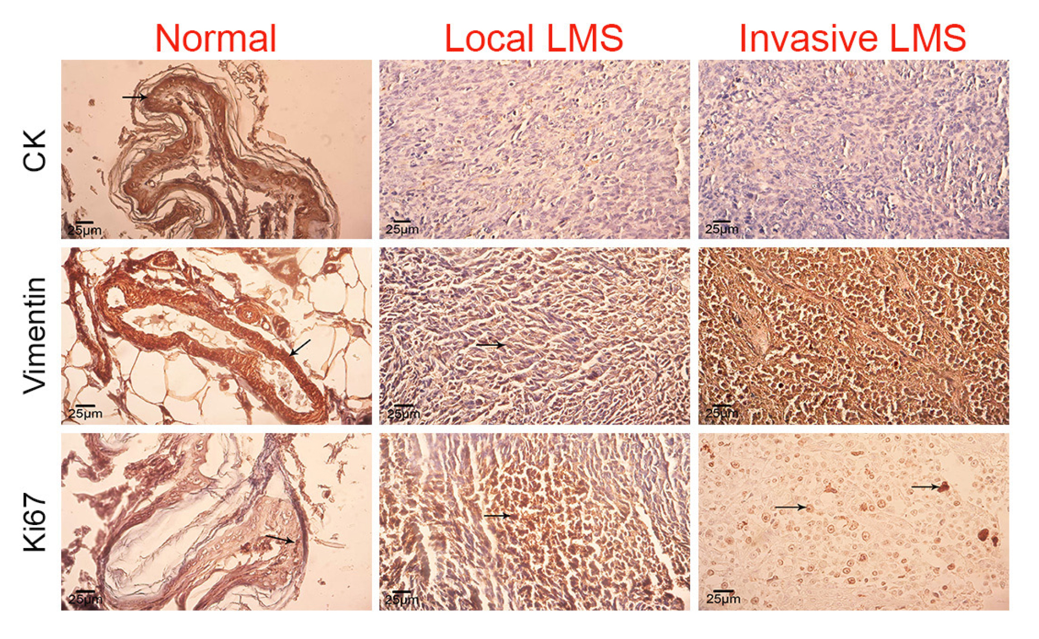
Figure 7.The following comparative immunohistochemical photomicrographs of normal skin tissue, local and invasive LMS. The strong CK expression in normal squamous skin cells, its weak expression in the local tumors, and lack of expression in the invasive tumor confirm the mesenchymal origin of the tumor cells. The strong Vimentin expression in the smooth muscles around the blood vessels (arrow), normal skin tissue, moderate cytoplasmic expression of Vimentin in the local tumor, and its strong and dispersed expression in the invasive tumor can be seen. A weak Ki67 expression is visible inside the nuclei of several normal squamous skin cells (arrow), in addition to a strong and dispersed expression inside the nuclei of local tumor cells (arrow), and a moderate and dispersed nuclear expression in the invasive tumor cells (arrow) (IHC).
Discussion
The present study investigated the occurrence, clinicopathological features, hematological alterations, and immunohistochemical characteristics of cutaneous leiomyosarcoma (LMS) in dogs.
Results of the study, conducted over 10 years in Urmia city, revealed that among the 318 dogs with various cutaneous lesions, 6 (1.88%) were diagnosed as LMS (local and invasive). According to the occurrence rate of this tumor in other countries, information from 748 cases of neoplasm in dogs in Korea, leiomyoma and leiomyosarcoma had 0.27% and 0.4% frequencies, respectively [ 8 ]. Similarly, retrospective research conducted over seven years on dog skin tumors in analytical research center, north region of Portugal (2014 to 2020), out of the 1185 cases with lesions identified as cutaneous neoplasms, 62.9% of them were reported as benign, and 37.1% as malignant. Mast cell tumors (22.7%) were the most common form of identified tumor, thereafter benign soft tissue neoplasms (9.7%), Sebaceous gland tumors (8.1%), vascular neoplasms (7.9%), and soft tissue sarcomas (7.6%). Cutaneous cancers were multicentric (6.14%), followed by solitary tumors in hind limb areas (12.1%), anterior motor organs (8.6%), buttocks (7.1%), abdomen (6.5%), and rib area (5.2%) [ 9 ]. In the current study, Malignant cutaneous cancers of smooth muscles affected dogs of both sexes, predominantly at high ages (between 6 and 12 years). Although two affected dogs belonged to small breeds, the majority, were medium to large breeds, supporting earlier suggestions observed in previous research [ 1 , 10 ], despite the fact that these tumors are rare, they are more common in larger dog breeds. On the other hand, some other studies also demonstrated that the occurrence of LMS tumors is not related to the dog's breed potential [ 11 ]. In the present study, hematological analyses showed significant decrease in indices related to red blood cells, such as Hct, Hb, RBC, and MCHC in dogs affected by LMS compared with healthy dogs (p < 0.05), indicating anemia. However, this anemia was more pronounced in invasive LMS group compared to local LMS group. In terms of white blood cell indices, total WBC, Mon, and Eos in LMS groups had a significant decrease compared with healthy dogs (p < 0.05), indicating the involvement of immune system in LMS groups with the tumor and possible existence of a chronic inflammation. In another study on 44 dogs (1983-1988) with leiomyosarcoma (spleen, stomach, small intestine, cecum, and liver), every dog with liver LMS was clearly involved with metastasis, and they were euthanized during the surgery. In the remaining three groups, 79% of the infected dogs had no indication of metastasis during the surgery. In this research abnormal hematologic and biochemical findings included leukocytosis (8 cases), anemia (7 cases), azotemia (4 cases), increased serum ALP levels (6 cases), and elevated serum ALT and AST levels (3 cases) [ 11 ]. The outcomes of the mentioned study were in agreement with the findings of the present study regarding the occurrence of leukocytosis and anemia. Although the recent study did not investigate the cutaneous LMS, symptoms including anemia was observed in local LMS case and invasive LMS cases, further supporting the presence of paraneoplastic syndromes, which are commonly associated with malignant neoplasms. Besides, one of the cases with local LMS and two cases with invasive LMS indicated hypoglycemia, which is also among the symptoms of paraneoplastic syndromes [ 12 ]. In the present study, all local LMS tumors were solitary, and the invasive LMS tumors were dispersed. The local LMSs were related to surface cutaneous tissues, and no recurrence observed during a six-month follow-up after the surgery. In contrast, among the invasive LMS cases, two animals were euthanized due to disease severity and metastatic nature of the disease, While follow-up data were unavailable for the remaining case due to owner refusal of euthanasia. These observations suggest that early surgical intervention in local LMS may result in a favorable prognosis. Previous studies have similarly reports an acceptable prognosis for cutaneous and subcutaneous smooth muscle tumors in dogs; however, such information is primarily limited to tumors originating from the smooth muscles that erect hair or from the walls of blood vessels, and there is no information on the smooth muscle tumors inside the deep soft tissues [ 13 ]. Immunohistochemistry evaluation for Myogenin, MyoD1, and CD31 showed that the negative results of LMSs for the mentioned markers confirmed their definite LMS nature and piloleiomyosarcoma type. These three markers were primarily intended for the differential diagnosis of LMS from other muscle tissue tumors, such as rhabdomyosarcoma, and the differential diagnosis of LMS types (piloleiomyosarcoma and angioleiomyosarcoma). In the present study, SMA and Desmin was significantly increased in LMS tissues compared with normal skin, although the extent of changes in their expression and intensity varied based on tumor's nature and biological behavior. Vimentin played a significant role in the metastasis process and was investigated under laboratory conditions. During the epithelial-mesenchymal transition (EMT), epithelial cells lose their basoapical and intercellular polarity, and obtain adhesion features by the downregulation of genes related to epithelial cells, including E-cadherin and cytokeratins, and while acquiring migrative and invasive characteristics related to mesenchymal cell phenotypes through genes such as N-cadherin and Vimentin [ 14 ]. In other words, the process of epithelial-mesenchymal transition (EMT) is characterized by the suppression of epithelial phenotypes and the concomitant adoption of mesenchymal properties. Cells in EMT frequently reside in a partial or intermediate state, co-expressing both epithelial and mesenchymal markers, with a fully mesenchymal state being uncommon. Notably, this transition is reversible through its counterpart process, mesenchymal-epithelial transition (MET) [ 15 ]. The progressive increase in Vimentin expression with increasing tumor metastatic aggressiveness observed in the present study supports its role as an indicator effective in the degree of malignancy or invasiveness of LMS. In a report related to the first primary leiomyosarcoma occurrence in the testicles of two 10 and 12 year old dogs, the immunohistochemistry findings demonstrated a positive (moderate) immune reaction by the tumor cells to Vimentin, SMA, and Desmin markers [ 16 ]. A report from Japan on multiple polymorphic cutaneous leiomyosarcomas in a 13-year-old male Shih Tzu dog described leiomyosarcomas with poor differentiation and explained that their distinction from anaplastic sarcomas with giant cells was difficult. Immunohistochemistry examination indicated the high positivity of tumor cells to Vimentin, their relative positivity to SMA and Desmin, and a negative response to cytokeratin [ 3 ]. In a systematic review of 2616 dogs and cats with skin tumors in Britain, the occurrence rate of cutaneous leiomyomas in dogs and cats was 0.88% and 0.33%, respectively. Differentiating leiomyosarcoma, rhabdomyosarcoma, and fibrosarcoma using routine histological methods might be difficult; however, ultrastructural and immunohistochemical methods are useful for the accurate diagnosis of soft tissue sarcoma tumors. A report from Korea described one cutaneous piloleiomyoma and two angioleiomyosarcoma cases in three female dogs aged 7 to 12 years with solitary or paired nodules. In terms of immunohistochemistry, tumor cells showed extremely positive reactions to SMA [ 8 ]. A retrospective study identified 24 dogs with histological diagnosis of definite or suspected leiomyoma and leiomyosarcoma in a non-visceral location. According to immunohistochemistry tests, more than two-thirds of the leiomyosarcoma tumor cells showed positive reactions to SMA and Laminin markers. Histochemistry also indicated a mild to moderate matrix deposition, which was identified using Masson's trichrome staining. The study data indicated the unusual nature of non-visceral leiomyosarcoma and the importance of IHC for their diagnosis [ 17 ]. In several malignant tumors related to different diseases, EGFR is overexpressed. By overexpressing these molecules in neoplasms, they will be involved in the functions of some critical stages, such as vascularization, inhibition of programmed cell death, immunosuppression, enhanced cellular multiplication, possibility of invasion, and cellular specialization and displacement. Due to the importance of EGFR and its involvement in the progression and malignancy, reduced survival rate, and poor prognosis, they are considered promising biomarker and potential therapeutic targets in veterinary oncology [ 7 ]. In a report related to a high-grade sarcoma case of an eleven-year-olds neutered female Labrador Retriever, immunohistochemistry analysis demonstrated the positive VEGFr, PDGFr, SCF, and EGFR in the tumor cells. Repeating the surgical removal and targeted treatment with toceranib led to a stable recovery for about two years. IHC results for the EGFR maker in the present study indicated a remarkable increase in the expression and intensity of this maker in the local LMS category in comparison with the normal and invasive LMS categories. Experimental studies transplanted highly tumorigenic and chemotherapy-resistant human LMS cells to mice and regenerated the tumor, and demonstrated that these cells indicated the triggering of EGFR/AKT/MAPK pathways, implying the potential of prevailing their drug resistance by blocking EGFR [ 18 ]. Moreover, TGFα-EGFR signaling pathways has been implicated in enhancing tumor growth rate and its metastasis. Moreover, the signaling activates the endothelial cells related to the tumor in addition to the tumor cells [ 19 ]. Although EGFR is specific to tumors with epithelial cell origin, the present study determined that EGFR expression occurs both in epithelial tumors and mesenchymal tumors, such as LMS, and as EGFR expression decreases in the invasive LMS cells, it may be an indicate that EGFR upregulation plays a role during early tumor development and its expression possibly continues until the metastasis stage. However, confirming this finding requires additional molecular analyses. Analysis of E-Cadherin and β-Catenin expression demonstrated that increased β-Catenin expression and intensity in local LMS cells compared with invasive LMS and normal skin cells was significantly higher than E-Cadherin, and although E-Cadherin expression in the local LMS group was higher than invasive LMS and normal skin, when applying a 10% threshold, the increase in E-Cadherin (under 10%) expression was negligible compared with β-Catenin (40% to 60%). Epithelial cells are characterized by the expression of E-Cadherin, which is conceptualized as both a tumor suppressor gene and a morphogenic factor within the context of epithelial neoplasms. Studies have indicated a reduction or absent in E-Cadherin expression correlating with a decrease in differentiation, invasion, or metastasis among various malignancies, including multiple forms of sarcoma. An investigation into the expression of E-Cadherin, β-Catenin, and topoisomerase IIα in human leiomyosarcoma analyzed 19 paraffin-embedded primary and non-metastatic leiomyosarcoma tissue specimens for the expression of these markers utilizing immunohistochemistry (IHC), employing a threshold of 20% for determining positive cell staining results. The outcomes of this study indicated that E-Cadherin expression was uniformly negative across all leiomyosarcoma samples. Additionally, negative β-Catenin nuclear expression was found in all leiomyosarcoma samples, whereas immunopositivity for cytoplasmic expression of β-Catenin was recorded in nearly 50% of the patients [ 20 ]. β-Catenin/E-Cadherin expression was not investigated in dogs; however, a study on the mentioned issue in canine papilloma and SCC skin tumors reported through immunohistochemistry analyses that inappropriate β-Catenin/E-Cadherin expression occurred during the epidermal tumorigenesis in dogs. Such results support the hypothesis indicating that inappropriate β-Catenin/E-Cadherin expression may has a crucial importance in the pathogenesis of canine epidermal neoplasms, and not merely due to the disruption of the intercellular junctions, but also because of the irregular activating of the signaling pathways that these molecules are involved with [ 21 ]. Considering the β-Catenin/E-Cadherin IHC results in the current study, it appears that β-Catenin and E-Cadherin alone may not be sufficient prognostic markers, but may contribute to understanding tumor invasion pathways [ 22 ].
Ki67 immunoreactivity in the present study was significantly increased in both invasive and local LMS groups compared with the normal animals. Hence, considering the malignant and invasive nature of the tumor, the cell proliferation rate was high, as the percentage of Ki67-positive cell nuclei was 60% to 80%, and subsequently, the H-Score of the tumor groups had a significant increase compared with the normal group. It must be noted that Ki67 expression in canine beta tumor cells is highly dependent on the tumor type, and other factors, such as the animal's age and sex, can somewhat play roles in changing the marker's expression in tumor tissues [ 23 ]. In humans, studies suggested labelling indices (LI) over 10% as a tumor prognosis index for uterine leiomyosarcoma tumors [ 24 ]. Nevertheless, considering the aforementioned factors, LI may vary in different studies; therefore tumor prognosis should not rely solely on Ki-67 and LI, but should also consider additional histopathological parameters such as nuclear atypia and mitotic index [ 25 ]. Although CK maker is primarily used to diagnose tumors of epithelial cell origin, regarding the IHC results for this marker in the present study, considering the existing reports indicating the inappropriate expression of cytokeratin in normal and neoplastic tissues with non-epithelial origin [ 26 ], this marker was added to the immunohistochemistry panel. In the present study, CK expression was confined to squamous epithelial cells in normal skin, while LMS tumors exhibited low expression (under 10%) and negligible amount of CK immunoreactivity.
In summary, among 318 dogs with cutaneous lesions, 6 cases of leiomyosarcoma (LMS) were confirmed, including 3 local and 3 invasive. Blood test showed significant increases in leukocytes, monocytes, and eosinophils (p < 0.05) in affected dogs. Microscopic analysis revealed tumor fibers intermingled with connective tissue and marked cell heterogeneity. Tumor cells had a mean mitotic index of 4.2 for local cases and 5.8 for invasive ones. Immunohistochemical tests showed no MyoD1 or Myogenin, confirming LMS, and the tumors lacked CD31, indicating they were piloleiomyosarcoma. Notably, tumor cells had higher positive expressions of Desmin, SMA, Vimentin, and Ki67 compared to normal skin. Additionally, local LMS showed increased EGFR and β-Catenin compared to invasive LMS and normal tissue. The study demonstrate that, EGFR expression is not limited to epithelial tumors but is also relevant in mesenchymal tumors, such as LMS. While, β-Catenin and E-Cadherin alone are insufficient for prognostic assessment, they may help in understanding invasion pathways and aid in treatment decision making.
Ethical approval
The study was performed following approval from the Research Ethics Committee of Islamic Azad University, Urmia Branch (approval code: IR.IAU.URMIA.REC.1403.148; September 8, 2024).
Animals
Formalin fixed tissue samples and paraffin embedded blocks of canine cutaneous tumor lesions were retrospectively collected from cases referred to the Veterinary Clinic of the Islamic Azad University, Urmia Branch, and private pet clinics in Urmia city over a 10 year period (January 2014 to January 2024). During this period, a total of 318 dogs of various breeds, presenting with various cutaneous lesions were examined. These included Rottweiler (n= 25), Shih Tzu (n= 21), Iraqi (Kurdish Mastiff) (n= 15), German Shepherd (n= 33), Terrier (n= 87), and Iranian mixed breeds (n= 137).
Pathologcal Examination
For cases suspected of skin tumors, tissue samples were obtained either by biopsy (from local tumors) or during necropsy (invasive and metastatic tumors) and archived for histopathological evaluation. In macroscopic examination, first, the cutaneous lesions suspected of tumors were examined (i.e., size, number, color, consistency, hemorrhage, location, and distribution of tumors), and all the other relevant animal characteristics were recorded. Tissue samples were fixed in 10% neutral buffered formalin, stained using the H&E method and investigated using an optical microscope. In cases suspected of connective tissue and smooth muscle tumors, Masson's trichrome staining was used for differential diagnosis and to confirm the tumor. After fixation tissue samples were cut into 0.5 × 0.5 × 1 cm sections, and processed using an Autotechnicon Tissue Processor. Samples underwent dehydration, clearing, and paraffin impregnation, followed by sectioning at 6 μm thickness using a micrometers Sections were routinely stained with Hematoxylin and Eosin (H&E) method was used to stain. In cases where the cutaneous tumor mass was suspected of sarcoma, the Masson's trichrome method was applied to confirm and differentiate the diagnosis of leiomyosarcoma from fibrosarcoma and other fibromatous lesions [ 27 ]. Diagnostic criteria for cutaneous LMS, included cellular and nuclear pleomorphism (atypia), number of nucleoli, number of mitotic figures, and the presence of necrosis and hemorrhage in the tumor tissue section. Additionally, the features of the fibers with varying connective tissue amounts were investigated [ 13 ].
Hematologcal Examination
Blood samples were collected from the jugular veins of six healthy dogs and six dogs diagnosed with cutaneous leiomyosarcoma tumors using 21-gauge needles into EDTA-containing tubes. Hematological parameters were analyzed using an Abbott Cell Dyn 3500 automated hematology analyzer.
Immunohistochemistry
Immunohistochemical staining was performed using the EnVision® + HRP dual system. Sections were dewaxed with xylene and rehydrated with ethanol, followed by incubation in hydrogen peroxide (3%) for 30 minutes to inhibit non-specific peroxidase activity. Heat-induced epitope retrieval was conducted using a 0.01 M citrate buffer (pH = 6.0) for 25 minutes, and slides were incubated with albumin bovine serum (5%) in Tris buffered saline (TBS) for half an hour to suppress non-specific staining. Afterward, the sections were washed with water and TBS and held at room temperature. In the last stage, the sections were incubated overnight at 4°C with the following primary: α-SMA (Dako, USA; 1:200), Ki67 (Dako, USA;1:100), β-catenin (Dako, USA;1:100), cytokeratin (Dako, USA; 1:50), VIM (Dako, USA;1:100), MyoD1 (ThermoFisher, clone 5.8a, 1:50), Myogenin (ThermoFisher, clone FSD, 1:800), Desmin (Dako, USA; 1:100), EGFR (Zymed Laboratories, San Francisco, California, USA; 1:50), E-cadherin (Invitrogen, Carlsbad, CA, USA; 1:50), and CD31 (Dako, USA; 1:200). Following primary antibody incubation, slides were washed with phosphate-buffered saline (PBS) and incubated with streptavidin-horseradish peroxidase (HRP) for 20 minutes. After another wash with the buffer, the slides were treated with diaminobenzidine for 10 minutes, with the sections being counterstained using Harris hematoxylin [ 28 ].
CD31, which is a marker for endothelial cells of blood vessels, was used to differentiate LMS types (piloleiomyosarcoma originating from the smooth muscles around the hairs, and angioleiomyosarcoma originating from the cutaneous vessels). MyoD1 and Myogenin were used to differentiate Rhabdomyosarcoma (RMS) from LMS. cytokeratin and vimentin were used to determine epithelial or mesenchymal origin of the tumor cells, Ki67 to evaluate cellular proliferation rate, and α-SMA to confirm smooth muscle fibers differentiation. EGFR, β-catenin, and E-cadherin were evaluated to investigate possible EMT changes and prognostic in LMS tumor cells [ 2 , 7 , 14 ]. In order to rank the expression intensity of immunohistochemical markers for each sample (normal and pathological), five randomly selected microscopic fields (each measuring 2.37 mm2) were evaluated at ×40 magnification. The number of positive cells, exhibiting positive brown immunoreactivity reaction, was counted and expressed as a percentage of the total cells. The histochemical score assessment (H-Score) was calculated to assess both staining intensity and distribution using the following formula:
In this formula, P indicates the percentage of positive cells (+, ++, and +++) inside the tissue, represented by P1, P2, and P3, respectively. The numerical scale for H-Score ranged between 0 and 300 [ 29 , 30 ].
Statistical Analysis
Statistical analyzed were performed using GraphPad Prism software (Version 9). Descriptive findings, including mean, standard deviation, and standard error, were calculated. The Shapiro-Wilk test was used for evaluating quantitative data normality. In the next step, considering the scoring data normality of the immunohistochemical percentage and H-Score data, differences among normal, local LMS, and invasive LMS groups, were analyzed using one-way analysis of variance (one-way ANOVA), followed by Tukey's post-hoc tests. Statistical significance was set at p < 0.05.
Authors' Contributions
F.H. and A.A. conceived and planned the experiments. F.H. and A.A. carried out the experiments. F.H. and A.A. contributed to sample preparation. A.A. contributed to the interpretation of the results. A.A. took the lead in writing the manuscript. Both authors contributed to the first draft, revising.
Acknowledgements
We would like to thank the TUMS Cancer Institute for preparing the immunohistochemical sections.
Conflict of interest
The authors declare that there is no conflict of interest.
Abbreviations-Cont'd
LMS: Leiomyosarcoma
α-SMA: α-Smooth Muscle Actin
EGFR: Epidermal Growth Factor Receptor
COX-2: Cyclooxygenase-2
CK: Cytokeratin
EDTA: Ethylenediaminetetraacetic Acid
HRP: Horseradish Peroxidase
TBS: Tris-Buffered Saline
ANOVA: Analysis of Variance
H-Score: Histochemical Score
HCT: Hematocrit
Hb: Hemoglobin
RBC: Red Blood Cell
MCV: Mean Corpuscular Volume
MCHC: Mean Corpuscular Hemoglobin Concentration
PLT: Platelet
WBC: White Blood Cells
Neut: Neutrophil
Lym: Lymphocyte
MON: Monocyte
EOS: Eosinophils
VEGFr: Vascular Endothelial Growth Factor receptor
PDGFr: Platelet-Derived Growth Factor receptor
SCF: Stem Cell Factor
IHC: Immunohistochemistry
AKT/MAPK: Protein Kinase B and Mitogen-Activated Protein Kinase
TGFα: Transforming Growth Factor-alpha
SD: Standard Deviation
References
- Torrigiani F, Pierini A, Lowe R, Simčič P, Lubas G. Soft tissue sarcoma in dogs: A treatment review and a novel approach using electrochemotherapy in a case series. Vet Comp Oncol. 2019;17(3):234–41.DOI
- Guisado FR, Castro PL. Piloleiomyosarcoma in cats: Histological and immunohistochemical features. Vet Pathol. 2022;59(1):57–62.DOI
- Aihara N, Sugiyama J, Baba H, Kamiie J. Multiple cutaneous pleomorphic leiomyosarcoma in a dog. J Vet Med Sci. 2019;81(11):1564–6.DOI
- Kim YJ, Jeon Y, Bae H, Shin SW, Cho Ar, Hyun J-E, et al. Oral leiomyosarcoma in a dog. J Biomed Transl Res. 2021;22(4):199–204.DOI
- Han J, Kim Y, Kim D, Na K. Alteration in E-Cadherin/b-Catenin expression in canine melanotic tumors. Vet Pathol. 2012;50(2):274–80.DOI
- Bongiovanni L, Malatesta D, Brachelente C, D'Egidio S, Della Salda L. β-Catenin in canine skin: Immunohistochemical pattern of expression in normal skin and cutaneous epithelial tumours. J Comp Pathol. 2011;145(2–3):138–47.DOI
- Files R, Santos C, Queiroga FL, Silva F, Delgado L, Pires I, et al. Investigating Cox-2 and EGFR as biomarkers in canine oral squamous cell carcinoma: Implications for diagnosis and therapy. Curr Issues Mol Biol. 2024;46:485–497.DOI
- Jung J-Y, Kang S-C, Park D-S, Lee E-S, Bae J-H, Kim J-H. Cutaneous smooth muscle tumors in 3 dogs. Korean J Vet Res. 2009;49(1):63–6.
- Martins AL, CanadasSousa A, Mesquita JR, DiasPereir P, Amorim I, Gärtner F. Retrospective study of canine cutaneous tumors submitted to a diagnostic pathology laboratory in Northern Portugal (2014–2020). Canine Med Genet. 2022;9(1):2.DOI
- Jacobs TM, Poehlmann CE, Kiupel M. Injection-site sarcoma in a dog: clinical and pathological findings. Case Reports Vet Med. 2017;2017:1–4.DOI
- Kapatkin AS, Mullen HS, Matthiesen DT, Patnaik AK. Leiomyosarcoma in dogs: 44 cases (1983-1988). JAVMA. 1992;201(7):1077–9.
- Meuten DJ. Tumors in Domestic Animals. Fifth. Wiley Blackwell; 2017. 15–16 p.
- Avallone G, Pellegrino V, Muscatello LV, Roccabianca P, Castellani G, Sala C, et al. Canine smooth muscle tumors: A clinicopathological study. Vet Pathol. 2022;59(2):244–55.DOI
- Berr AL, Wiese K, Santos G dos, Koch CM, Anekalla KR, Kidd M, et al. Vimentin is required for tumor progression and metastasis in a mouse model of non–small cell lung cancer. Oncogene. 2023;42:2074–8.DOI
- Yim IS, Laronde DM. Biomarkers of epithelial-mesenchymal transition: E-cadherin and beta-catenin in malignant transformation of oral lesions. Can J Dent Hyg CJDH = J Can l'hygiene Dent JCHD. 2024;58(2):111–9.
- Ciaputa R, Brambilla E, Godizzi F, Dzimira S, Żebrowski K, Kandefer-Gola M, et al. First report of primary testicular leiomyosarcoma in two dogs. BMC Vet Res. 2023;19:101.DOI
- Brady R V., Rebhun RB, Skorupski KA, Burton JH, Al-Nadaf S, Choi E, et al. Retrospective immunohistochemical investigation of suspected non-visceral leiomyosarcoma in dogs. J Vet Diagnostic Investig. 2022;34(3):465–73.DOI
- Sette G, Salvati V, Memeo L, Fecchi K, Colarossi C, Di PM, et al. EGFR Inhibition Abrogates Leiomyosarcoma Cell Chemoresistance through Inactivation of Survival Pathways and Impairment of CSC Potential. PLoS One. 2012;7(10):e46891.DOI
- Sasaki T, Hiroki K, Yamashita Y. The Role of Epidermal Growth Factor Receptor in Cancer Metastasis and Microenvironment. Biomed Res Int. 2013;2013:1–8.DOI
- Gogou PN, Batistatou A, Pakos EE, Apostolikas N, Stefanou D, Tsekeris PG. Expression of E-cadherin, ß-catenin and topoisomerase IIα in leiomyosarcomas. Clin Transl Oncol. 2009;11:548–51.DOI
- Sanz Ressel B, Massone A, Barbeito C. Aberrant expression of E-cadherin/b-catenin during epidermal tumourigenesis in dogs. J Comp Pathol. 2020;176:1–9.DOI
- Debruyne P, Vermeulen S, Mareel M. The role of the E-cadherin/catenin complex in gastrointestinal cancer. Acta Gastroenterol Belg. 1999;62:293–402.
- Firsching TC, Dietert K, Bartel A, Gruber MGD, D A. Dependence of the Ki67 Labelling Index of Selected Canine Tumours on Patient Age, Sex and Tumour Size. J Comp Pathol. 2022;193:1–8.DOI
- Travaglino A, Raffone A, Catena U, Luca M De, Toscano P, Prete E Del, et al. Ki67 as a prognostic marker in uterine leiomyosarcoma: A quantitative systematic review. Eur J Obstet Gynecol Reprod Biol. 2021;261:119–24.DOI
- Bergin IL, Smedley RC, Esplin DG, Spangler WL, Kiupel M. Prognostic Evaluation of Ki67 Threshold Value in Canine Oral Melanoma. Vet Pathol. 2011;48(1):41–53.DOI
- Adegboyega PA, Qiu S. Immunohistochemical profiling of cytokeratin expression by endometrial stroma sarcoma. Hum Pathol. 2008;39:1459–64.DOI
- Amoorahim S, Amniattalab A. Immunohistochemical expression of GDNF, P53 and Ki67 with Tunel assay in canine non-neoplastic esophageal nodules induced by Spirocerca lupi. Maced Vet Rev. 2025;48(1):39–51.DOI
- Babanezhad-gajouti A, Amniattalab A. in the canine spleen with siderofibrotic nodules : an immunohistochemical and immunofluorescence perspective on apoptosis , ferroptosis , and oxidative stress. Turkish J Vet Anim Sci. 2023;47(4):397–412.DOI
- Piercealla WE, Wolfea M, Suschaka J, Changa H, Chena Y, Sprotta KM, et al. Strategies for H-score normalization of preanalytical technical variables with potential utility to immunohistochemical-based biomarker quantitation in therapeutic response diagnostics. Anal Cell Pathol. 2011;34:159–68.DOI
- Ruengwanichayakun P. Histochemical scoring assessment (H-score). Asian Arch Pathol. 2021;3(1):13–4.


