
Abbreviations
CEH: Cystic Endometrial Hyperplasia
DLH: Domestic Long Hair
DSH: Domestic Short Hair
Introduction
Pathological lesions of the uterus and ovaries, including the cystic endometrial/pyometrial hyperplasia complex, are most commonly reported in older cats, however, many lesions cannot be identified due to their subclinical nature. In addition to physical examination and diagnostic tests, histopathological evaluations following ovariohysterectomy plays a critical role in the accurate identification of uterine and ovarian lesions in cats. Subclinical uterine disorders such as pyometra, may turn into life-threatening conditions. Pyometra in both dogs and cats is frequently associated with cystic endometrial hyperplasia (CEH), a condition influenced by progesterone hormone imbalances. Under the hormonal effect, myometrial contractility decreases, while the growth of endometrial glands increase [ 1 ].
Histopathological evaluation, in conjunction with clinical findings, is a valuable diagnostic tool that enables the differentiation between uterine infections and other non-infections uterine disorders. Removal of the uterus and ovaries allows for histopathological examination of various diseases of the female reproductive system [ 2 ]. It is probably influenced by repeated exposure of the endometrium to high concentrations of estrogen in the luteal phase, although the cause of cystic hyperplasia of the endometrium is not completely clear. The immobility of the uterus and the secretions of the endometrial glands provide a suitable environment for the growth and proliferation of bacteria. Among intrauterine bacterium Escherichia coli is the most common isolated in pyometra [ 3 ].Clinically, most cats with pyometra exhibit signs such as anorexia, lethargy, weight loss, or even septic shock and peritonitis. In cats with open cervix pyometra a wet perineal area and purulent vaginal secretions are commonly observed, polyuria and polydipsia may also occur [ 4 ]. A reliable method to definitively diagnose pyometra is to evaluate the uterine by ultrasonography. In healthy cats, the uterine horn diameter usually ranges from 0.4 to 0.7 cm, depending on the stage of the estrous cycle. Appropriate treatment for pyometra is ovariohysterectomy; however, fluid therapy, antibiotic administration, and appetite stimulants are required before surgery to stabilize the patient's body. Evaluation of ovarian pathology is helpful in cases suspected of ovarian cystic hyperplasia [ 5 ]. Using dopamine agonist in medical treatment may delay the recurrences of pyometra for in the short term, however ovariohysterectomy remains the most effective and definitive treatment for pyometra [ 3 ].
Despite numerous studies conducted on feline reproductive disorders in recent years, the actual prevalence of uterine infection and their underlying mechanisms remain poorly understood [ 6 , 7 ]. In the USA and Europe,the proportion of privately owned cats that are neutered ranges from 27% to 93%. Regarding the frequency of histopathological lesions of the uterus and ovaries, there are relatively few reports in clinically healthy cats, with most available studies presented as isolated case reports. For instance, Pasteurella multocida was isolated from a 16-year-old Persian cat with pyometra, which presented with intermittent vomiting, anorexia and progressive abdominal distension [ 9 ]. In another survey involving 44 cats with inflammatory uterine diseases, ovarian cystic or active follicles were observed in 19 cats; while the remaining 25 cats exhibited luteal phase ovaries [ 10 ] Breed predisposition has also been reported, with a higher prevalence of pyometra in Bengal cats, accompanied by active corpora lutea on the ovaries, in connection with proliferative adenomatous changes, in the glandular and surface epithelium of uterine [ 11 ].
In another study conducted on fourty five cats with pyometra; it was showed that although dopamine agonist therapy may temporarily delay disease recurrence; ovariohysterectomy remains the most effective treatment option [ 5 ].
In Iran Considering that ovariohysterectomy is not routinely performed in privately owned cats articularly those kept individually. As a result, the prevalence of uterine infections in the domestic cats population may be relatively high. To date, there was no comprehensive study investigating uterine and ovarian pathologies in cats in Iran. The present study was conducted for the first time in cats of Ahvaz district in Khuzestan province. The current survey is practical and the obtained results will be of great help to veterinary clinicians. The purpose of the present study was to evaluate the histopathological lesions of the uterus and ovaries, including cystic hyperplasia of the uterus, pyometra, mucometra, neoplastic conditions in cats. Additionally, this study aimed to investigate the clinical signs and emphasizes the diagnostic and preventive value of ovariohysterectomy in the feline population in Ahvaz district.
Results
Clinical findings
In the present study, a total of fifty-four cats were evaluated. Among them, three cats were pregnanent, two cats had blood-filled uterus, and the remaining cats showed normal uterine and ovarian structures. The mean±SD of the studied cats were 10.94 ± 0.74 months, with an age range from six months to 2.5 years.
No significant difference association was observed between access to outdoor environments and the presence of uterine or ovarian lesions (p > 0.05). Only five cats had access to outdoor envirements. There was no recorded history of uterine diseases and administration of different drugs, including corticosteroids. At the time of examination, clinical findings, including vulva and vagina condition, presence of vaginal discharge, mammary gland abnormalities, lactation status, polyuria, polydipsia, and skin and hair coat condition, were within normal limits in all cats.
Based on the microscopic evaluation of the uterus and ovaries, cats were classified into four groups according to the estrous cycle:
Proestrus (Group 1): twenty-one cats were in the proestrous phase, In these samples, several large follicles were observed in the ovary and corpus luteum. Also, the uterine endometrium was thin and mildly hyperemic.
Estrus (Group 2): Fourteen cats were classified as being in estrus cycle. In these samples, the uterine mucosa showed marked hyperemia and superficial hemorrhage, with a large number of abundant erythrocytes present within the endometrial layer. A very large follicle was observed in the ovary.
Diestrus (Group 3): Eight cats were in diestrous phase. A large corpus luteum was present in the ovaries, and the endometrial layer was markedly thickened due to the proliferation of endometrial glandular cells.
Anestrus (Group 4): Eleven cats classified as anestrous. In these samples, ovarian follicles exhibited the smallest diameter, and the corpora lutea was markedly regressed.
Among thirteen Persian cat, five were in proestrus phase, three in estrus, four in anestrus, and one in diestrus. Thirty-nine cats were of the DSH breed; among them, fourteen were in proestrous, ten in estrous, eight in diestrous in all cats, and seven in anestrous. One DLH cat and one Siamese cat were in the proestrus phase. They were showing irregularities in estrous cycle distribution, which may be attributed to induced ovulation in cats or differences in environmental and management conditions [ 3 ].
Histopathological examination revealed uterine or ovarian lesions in five out of fifty-four cats, Identified lesions included: one case of endometrial cystic hyperplasia, one case of granulosa cell tumor, one case of uterine cyst with ovarian hemorrhage, one case of uterine and ovarian hyperemia with hemorrhage, and one case of uterine hemosiderosis.
Out of five affected cats, involvement was observed in four cases (80%), ovarian involvement in two cases (40%), and simultaneous uterine and ovarian involvement in two cases (40%). All lesions were identified in DSH breed. The mean±SD age of affected DSH cats was 10.71±0.95 months. The relatively high standard deviation may be related to variations in age at ovariohysterectomy and the small number of affected cases[ 12 ].
Measurements of endometrial thickness, myometrial thickness, total uterine thickness, the number of follicles and number of corpora lutea were performed in all samples. The fifty-four uterine and ovarian samples were categorized into four estrous cycle groups. In the diestrous group (Group 3), a reduction was observed in endometrial thickness, myometrial thickness, uterine thickness, and the number of large corpora lutea. The greatest amount of thickness was recorded in the second group, during the estrous phase (p < 0.01; Table 1). The number of follicles gradually decreased from Group 2 to Group 4 (p < 0.05). The highest mean ± SD number of follicles was observed in the proestrous group (11.76 ± 0.95), with a maximum of 17 follicles. The lowest follicles count was noted in the cat affected by CEH. Although variations were observed in cats with uterine cysts and ovarian hemorrhages, these differencse were not sataistically significant. In the estrous group, the maximum of uterine thickness was 15.11 ± 0.78 µm, while the lowest uterine thickness (6.5 µm) was observed in the cat affected by granulosa cell tumor, possibly due to the effect of the tumor in uterus (p < 0.05). No significant difference were observed in the number of corpora lutea among the four groups in the involved samples.
| Mean ± SD | Cycle | |||
|---|---|---|---|---|
| Proestrus | Estrus | Diestrus | Anestrus | |
| 9.21 ± 0.93 | 11.00 ± 1.21 | 11.14 ± 0.61 | 9.33 ± 0.64 | Tickness of endometrium |
| 1.43 ± 0.25 | 1.55 ± 0.07 | 1.84 ± 0.16 | 1.49 ± 0.16 | Tickness of myometrium |
| 13.48 ± 0.94 | 14.75 ± 0.99 | 15.11 ± 0.78 | 13.79 ± 0.59 | Tickness of uterine |
| 10.66 ± 0.66 | 7.00 ± 0.90 | 11.42 ± 0.97 | 11.76 ± 0.95 | Number of follicles |
| 1.05 ± 0.34 | 2.42 ± 0.52 | 2.87 ± 0.58 | 2.66 ± 0.68 | Number of corpus luteum |
Among the five cats affected by uterine and ovarian disease, corpora lutea were observed in four cases. Two cats were in the proestrous cycle, one in estrous and two in diestrous. Microscopic examination revealed pathological changes in both uterus and ovaries tissues. One cat was diagnosed with cystic endometrial hyperplasia. This sample showed proliferation of endometrial cells with cysts formation within the endometrial mucosa. Additionally, several large follicles and corpora lutea were observed in the ovary. This lesion was categorized as Grade I CEH, based on the Dow classification (1962) [ 11 , 13 ] (Figures 1 and 2).
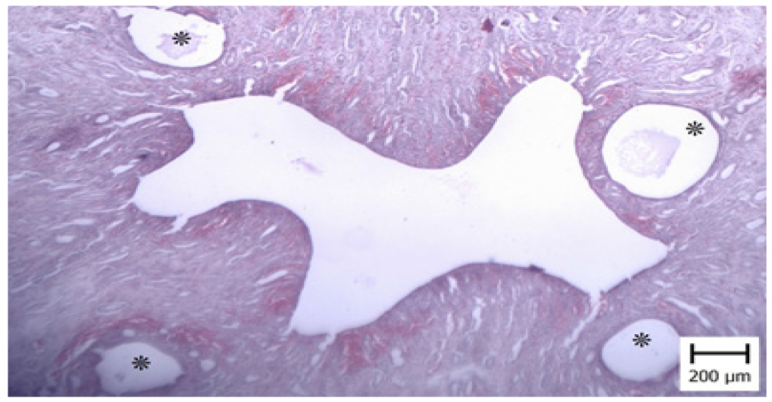
Figure 1. Cystic hyperplasia of the uterine endometrium.Note on cysts of varying sizes (asterisks) (Hematoxylin and Eosin).
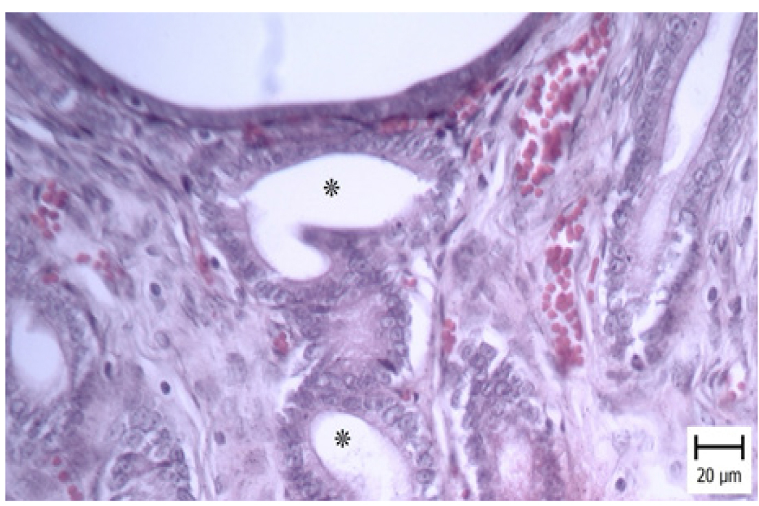
Figure 2. Higher magnification of the previous figure. Note proliferating columnar cells of the endometrial glands (asterisks) (Hematoxylin and Eosin).
In one case, severe hemorrhage was observed in the uterine perimetrium and ovary. A large ovarian hematoma containing numerous erythrocytes was detected in ovary (Figures 3 and 4).
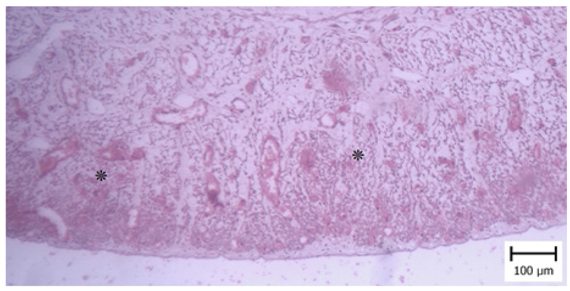
Figure 3. Hemorrhage (asterisks) in the uterine perimetrium (Hematoxylin and Eosin).
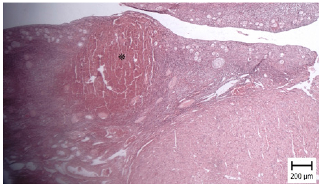
Figure 4. Hematoma in the ovary. Note to the accumulation of erythrocytes (asterisk) at the periphery of ovary (Hematoxylin and Eosin).
In another case diagnosed with hemosiderosis, iron particles were observed within uterine cells. In the sample with uterine cyst and ovarian hyperemia, hematoma and accumulation of erythrocytes were observed, consistent with severe uterine and ovarian hemorrhage. Microscopic evaluation of the tumor affected samples, revealed a mass of proliferating cells in ovarian tissue, arranged in the form of ladder and ropes. Minimal connective tissue and blood vessels were observed at the periphery of the proliferated cells layer. Tumor cells had large nuclei with one to two distinct nucleoli and a small amount of cytoplasm around them. Based on microscopic features, the lesion was diagnosed as a granulosa cell tumor (Figures 5 and 6).
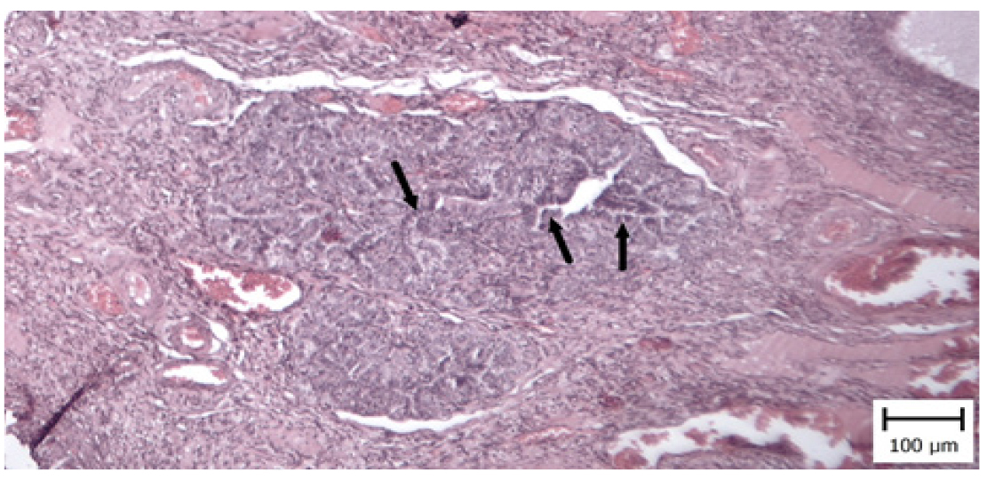
Figure 5. Granulosa cell tumor in the ovary. Note on foci of proliferated columnar cells (arrows) within the ovarian tissue (Hematoxylin and Eosin).
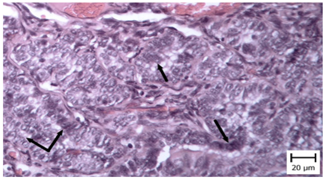
Figure 6. Granulosa cell tumor in the ovary. A higher magnification of the previous image. Note proliferating columnar with vesicular nuclei arranged in a palisading pattern (arrows) (Hematoxylin and Eosin).
Discussion
The aim of the present study was to evaluate histopathologyical lesions of the uterus and ovaries and emphasizes the diagnostic role of ovariohysterectomy in cats from Ahvaz region.There are relatively few reports on the frequency of histopathological lesions of uterine and ovarian in cats, and most available publications are limited to case report. For example, in a study on pyometra in a 16-year-old Persian cat, from which Pasteurella multocida was isolated. The disease in that case was started from two years ago, with symptoms intermittent vomiting, anorexia and progressive abdominal distension, pallor of mucous membranes, tachycardia, tachypnea, poor hair coat condition and severe emaciation [ 9 ]. In contrast, all fifty-four cats included in the present study were clinically healthy and showed no apparent clinical signs.
In another study, conducted on sixty dogs with cystic endometrial hyperplasia and pyometra, eighteen cases were diagnosed with open pyometra and three cases with closed form. Histopathological examination revealed hyperplasia of the endometrial glandular epithelium accompanied by numerous cysts [ 14 ]. Similarly, in the present study, the cat diagnosed with endometrial cystic hyperplasia showed proliferated of endometrial cells and cysts formation within the endometrial mucosa. According to the Dow classification (1962), this lesion was categorized as Grade I CEH [ 13 ]. However, unlike the canine study, no cases of pyometra were identified in the present study. The difference between two studies may be attributed to the higher incidence of pyometra in dogs compared with cats [ 3 ].
CEH is not associated with any specific phase of the estrous cycle; however, uterine enlargement is usually observed in affected animals [ 15 ]. in the present study, morphometric measurements of uterine thickness showed no significant difference between healthy cats and the cat affected by CEH. This finding may be explained by the early stage or short duration of the disorder in the affected animal [ 3 ]. Additionally, the small number of affected cases and young age of of the cats limit definitive conclusions and do not allow for direct comparison with other studies.
CEH is considered relatively common in cats and is characterized by endometrial epithelial hyperplasia and cystic dilation of endometrial glands. Affected uteri may appear covered with translucent cysts. Cats with CEH usually do not show clinical signs unless the disease is complicated by infection. Vaginal bleeding is rare may occur in association with uterine or ovarian torsion, caused by polypoid changes [ 15 ]. In the present study, only one case of the grade I CEH was detected among fifty-four samples (based on the Dow grading, 1962) [ 13 ], and no vaginal bleeding and uterine torsion was observed in this sample. Except FOR two cases with uterine hyperemia, all other uteri and ovaries appeared to be in normal status. Pyometra is regarded as one of the most important uterine disease in cats, develops secondary to CEH. The incidence of CEH increases with age, possibly due to cumulative nature of the disease. One of the definitive methods in the treatment of this disease is ovariohysterectomy. The cause of pyometra is complex and probably depends on various factors; but hormonal influences play a critical role in the pathogenesis of pyometra [ 16 ]. In the present study, the affected cat by CEH was two years and three months old, indicating that CEH can occur at a relatively young age, even before three years.
In a case report, on an affected dog to pyometra, abdominal distension was observed due to the accumulation of the purulent fluids, which had led to pressure on the rectum and intestinal obstruction. In histopathological confirmation, the remaining white tissue was ovarian tissue remnants [ 17 ]. In the present study, none of the cats were referred after ovariohysterectomy. Previous research has demonstrated associations between age, breed and the occurrence of pyometra in cats. Breeds such as Siberian, Osikat, Kurat, Siamese, Ragdoll, Maine Coon and Bengal are susceptible to this disease. The average age of pyometra in cats has been reported to be approximately four years, with a mortality rate of 5.7% slightly higher than existing reports in dogs (3-4%) [ 11 ]. In the present study, only uterine and ovarian lesions were observed in DSH cats, which may be related to the higher representation of this breed in the study population.
In another study involving seventy-nine female cats with endometrial hyperplasia with or without pyometra, uterine lesions were more prevalent in cats older than five years. Reproductive history and age at first pregnancy had no significant relationship with pathological symptoms of the disease. Microscopic evidence of pyometra was observed in 71% of clinically symptomatic cats, compared with only 19% of asymptomatic cats. These findings suggest an increased risk with advancing age. [ 18 ]; in the present study, the age of the affected cat was two years and three months, indicating that CEH also can develop even before age of three years.
In a study, a 12-year-old, sexually intact, female DSH cat with mild weight loss and recurring vaginal discharge, was diagnosed with CHE, accumulation of mucoid fluid in lumen of uterine corpus, uterine corpus adenocarcinoma and uterine cervix leiomyoma, following histopathological and immunohistochemical examination. [ 19 ].
Additionally, in a study examining histopathological features of endometrial lesions in six cats, one cat with papillomatous nodules and three cats with pyometra were identified. According to their results, the distribution of endometrial histopathological changes in cats were similar to humans [ 20 ]. In the present study, irregular proliferated of endometrial cells was observed in the cat affected by CEH.
A large-scale study involving 106 cats reported pathological alterations of the uterus and ovaries in twenty-nine cats. Ovarian cysts were found in 15 cats. Six cats had purulent endometritis with or without distension of the uterine lumen. Hyperplastic lesions of the endometrium were detected in two cats. In one cat, a uterine horn malformation with duplication of one uterine horn lumen was diagnosed. In their study, the prevalence of lesions increased with age, and fifty percent of female cats older than 3 years and eighty-three percent of cats over 5 years showed more or less pronounced pathological alterations of the uterus or ovaries. This may indicate a cumulative effect of repeated gonadal steroid hormone stimulation in subsequent oestrous cycles [ 8 ]. The broader range of pathological lesions in that study compared with the present study is probably attributable to the larger sample size.
In a PCR-based study, uterine samples were collected from 50 cats, and Chlamydia. felis infection was detected in 6% of the samples. Non-specific histopathological changes included varying degrees of edema, hyperemia, hemorrhage, inflammation, necrosis, fibrosis, cyst formation and endometrial hyperplasia.
These results highlighted the potential role of C. felis in uterine pathology and underline the need for surveillance of this zoonotic agent in domestic cats [ 21 ]. Other studies have reported a significant level of fungal organisms in the feline reproductive systems [ 22 ]. Gelberg and McEntee (1985) [ 23 ] identified hyperplastic endometrial polyps in fourteen cats, and ten of them were associated with diffuse or multifocal CEH. Their morphological evidence showed that endometrial polyps arise from focal areas of CEH. No breed association was observed and polyps were more common in adult cats. In the present study, considering the low age mean of the samples, no morphological evidence of polyps in the focal areas of cystic hyperplasia were observed, possibly due to the relatively young age of the affected cat.
Regarding ovarian neoplasms, a study examining reproductive disorders in 28 female cats, aging between seven months to thirteen years, reported a prevalence of 35.29% for ovarian neoplasms and 5.88% for theca granulosa cell tumor [ 24 ]. In contrast, only one case of granulosa cells tumor was identified in the present study.
Previous case reports have described granulosa cell tumors in cats presenting with systemic and reproductive signs, including weakness, anorexia, weight loss, lethargy, vomiting, and estrous abnormalities despite ovariohysterectomy, with Persian cat suggested as a susceptible breed [ 25 , 26 ]. In the present study, the affected cat was seven months year old DSH cat with no clinical signs, suggesting early-stage disease, and probability of occurrence of this tumor in other breeds and young ages. The granulosa cell tumor in the present study was detected incidentally during histopathological examination.
Granulosa cell tumors are the most common type of ovarian tumor in cats, and are considered malignant, with potential for metastasis [ 25 ]. Histopathological findings in another study done by Tawfik et al., (2015) [ 24 ], have included vascular congestion, hemorrhage, and hemosiderin deposition of golden yellow to brown granules in uterine tissue. Uterine tumours are rare in cats, accounting for approximately 0.3% of all feline neoplasms, with endometrial adenocarcinoma and uterine leiomyoma being the most common tumours of the feline genital tract. Hyperplastic lesions are potentially associated with hormonal stimulation are more likely [ 26 ]. In the present study, iron particles deposition (hemosiderosis) was also observed in uterine cells; however, due to early ovariohysterectomy, assessment of metastatic behavior was not possible.
Ovarian hematoma is more common in maresbut are rarely reported in cats. They typicall result from excessive bleeding following ovulation in follicular cavity. Hematomas can be large (up to 10 cm) and may resolve spontaneously, and their rupture and drainage can result in affected animal’s death (3). In the present study, a large hematoma with extensive hemorrhage was observed in the uterus and ovary. No comprehensive study has been conducted on hematoma in the ovary of cats. This article provides valuable information on the histopathological findings in the uterus and ovaries of cats undergoing ovariohysterectomy in the Ahvaz district. One limitations of the present study was the relatively small sample size (fourty-four samples) and limited age range of the cats examined, all of which were collected over cases referred to Ahvaz Veterinary Hospital during approximately six months.
Conclusion
Based on the findings of the present study, histopathological evaluation plays a crucial role in controlling, managing and providing appropriate diagnostic views for veterinarians, particularly when clinical and gross examinations fail to reveal abnormalities. Histopathological findings identified uterine and ovarian lesions in five cats, including one case of cystic endometrial hyperplasia, one granulosa cell tumor, one uterine cyst with ovarian hemorrhage, one case of uterine and ovarian hyperemia with hemorrhage; and another one case of uterine hemosiderosis. Overall, the present study demonstrates that ovariohysterectomy in cats can play an important role in diagnosing pathological lesions of the uterus and ovaries, prevention and treatment of genital diseases. Further studies are recommended on a larger population of cats in other regions of Iran.
Declaration of Generative AI and AI-assisted technologies in the writing process
The research and writing were done wiyhout relying on the artificial intelligenca tools.
Ethical Approval
Ethical approval of this study was obtained from the Ethics Committee of Shahid Chamran University of Ahvaz (EE/1401.2.24.173448/scu.ac.ir).
In the present study, samples were collected from fifty-four cat, referred for ovariohysterectomy. Of these cats, 39 were domestic short hair (DSH), 13 were Persian, one was domestic long hair (DLH), and one was Siamese. The mean±SE age of the cats was 10.94 ± 0.74 months, with an age range of six months to 2.5 years. Body weight ranged from 2.1 to 4.2 kg (3.1 ± 0.45). The cats were referred to the Veterinary Hospital of Shahid Chamran University of Ahvaz or different clinics in Ahvaz city between October and February 1401. All cats were in a healthy condition, with no recorded history of underlying disease or administration of specific medications. One litter had more than one birth and three litters had a history of one birth. All four litters belonged to the DSH breeds and no pregnancy history was recorded in other breeds. Most cats were kept indoors, and only five cats had access to the outdoor environments. .
Before ovariohysterectomy, a detailed clinical history was recorded for each cat, including age, breed, for each cat, including, status of uterine infections, administration of various drugs (including corticosteroids), and clinical findings. Clinical examinations assessed the condition of the vulva and vagina, presence of vaginal purulent discharge, mammary gland abnormalities, lactating state, polyuria and polydipsia, and body coat condition.
Following ovariohysterectomy, the macroscopic evaluation of the ovary and uterus was performed to assess the presence and the type of secretions, as well as the number and size of any macroscopic cysts. For histopathology evaluation, uterine and ovarian tissue samples were fixed in 10% buffered formalin. After fixation, tissues were trimmed into smaller sections and placed under running water for tissue passage. The working steps were including dehydration, clarification and impregnation. Tissue sample were slowly embedded in paraffin blocks using standard metal molds, and the blocks were cooled on ice. A microtome machine was used for cutting and sections with a thickness of 2-3 microns. Prepared slides were stained with Hematoxylin and Eosin in the following standard protocols. For dehydration, slides were placed in 80% alcohol for 10 seconds and twice in 96% alcohol in three steps. In the next step, in order to clarify of the tissues, the slides were placed in two different containers in Gezilol. Finally, the slides were mounted using Montalan glue and examined with an optical microscope. In histological examination, samples were evaluated to determine the estrus cycle stage based on uterine and ovarian histological characteristics. Measurements included uterine wall thickness, endometrium and myometrium thickness, number of mature follicles, number of corpora lutea and follicles. All measurements and counts were performed using a graduated lens.
Grouping
After examining the prepared samples with a microscope based on the histological changes in the uterus (including congestion and hyperemia) and ovary (follicular development and corpus luteum condition), samples were classified into four groups (proestrous, estrus, diestrus and anestrus).
Statistical Analysis
This study was designed as a descriptive analysis. Data were expressed as percentages, and Mean±SD. Statistical analyses were performed using SPSS software, version 22 (SPSS Inc., IL, Chicago, USA).
Authors' Contributions
B.M., A.R., and Be.M. conceived and planned the research. S.G., and Sh.G. carried out the investigation. B.M., and Sh.G. contributed to sample preparation. B.M., A.R., and S.G. contributed to the interpretation of the results.
Acknowledgements
We would like to thank their gratitude to the research council of Shahid Chamran University of Ahvaz for financial support.
Conflict of interest
Authors declare that they have no conflict of interest. There were no ethical considerations to be considered in this research.
References
- Noakes DE, Parkinson TJ, England GCW. Infertility in the Bitch and Queen. In: Veterinary Reproduction and Obstetrics. 10th ed. Elsevier Co. Ltd; 2019. p. 593-612.DOI
- Chatdarong K, Rungsipipat A, Axner E, Linde, FC. Hysterographic appearance and uterine histology at different stages of the reproductive cycle and after progestagen treatment in the domestic cat. Theriogenology. 2004;64(1):12-29.DOI
- Ettinger SJ, Feldman EC. Textbook of Veterinary Internal Medicine. Diseases of the dog and cat. Vol. 2. Sixth ed. Saunders Elsevier, St. Louis, Missouri; 2017. P. 1646-90.
- Johnston SD, Root-Kustritz MV, Olson PN. Canine and Feline Theriogenology. W.B. Saunders Company, Philadelphia; 2001. p. 389-474.
- Misk TN, EL-sherry TM. Pyometra in Cats: Medical versus surgical treatment. J Curr Vet Res. 2020;2(1):82-8.DOI
- Agudelo CF. Cystic Endometrial Hyperplasia – Pyometra Complex in Cats. A review. Vet. Q. 2005;27(4):173-83.PubMed
- Ortega-Pacheco A, Gutiérrez-Blanco E, Jiménez-Coello M. Common lesions in the female reproductive tract of dogs and cats. Vet Clin North Am Small Anim Pract. 2012;42:547-59.DOI
- Binder Cl, Reifinger M, Aurich J, Aurich Ch. Histopathological findings in the uteri and ovaries of clinically healthy cats presented for routine spaying. J Feline Med Surg. 2021;23(8):770-776.DOI
- Ashrafi Helan J, Hashemi Asal MM, Vajhi A, Shirani D, Fatahian HR, Mohit Mafi S, et al. (2002). Clinical report of a case of endometrial cystic hyperplasia/ pyometra grade III in a dog. Iran J Vet Res. 2002;4(1):94-101.
- Lawler DF, Evans RH, Reimers TJ, Colby ED, Monti KL. Histopathologic features, environmental factors, and serum estrogen, progesterone, and prolactin values associated with ovarian phase and inflammatory uterine disease in cats. Am J Vet Res. 1991;52(10):1747-53.PubMed
- Hagman R, Holst BS, Moller L, Egenvall A. Incidence of pyometra in Swedish insured cats. Theriogenology. 2014; 82(1):114-20.DOI
- Thrusfield M, Christley R, Brown H, Diggle PJ, French N, Howe K, et al., Veterinary Epidemiology. ath ed. john Wiley. Sons Ltd:2018. p.275-77.
- Dow C. The cystic hyperplasia-pyometra complex in the bitch. J Comp Pathol. 1959; 74:141-7.DOI
- Khalaf Deris S, Mosallanejad B, Rezaei A, Razi Jalali M, Ronagh A, Gooraninejad S. Evaluation of the clinical, hematological, biochemical and histopathological finding in bitches suffering from cystic endometrial hyperplasia/pyometra. Iran Vet J. 2022:18(5):1-12.
- Axner E. Clinical approach to conditions of the non-pregnant and neutered Queen. In: BSAVA Manual of Canine and Feline Reproduction and Neonatology, 2nd Ed, ed. England, G., & Von Heimendahl, A, British Small Animal Veterinary Association, Gloucester. 2010. P. 185-190.DOI
- dos Anjos Pires M, Vilhena H, Miranda S, Pereira MT, Seixas F, Saraiva AL. Proliferative endometrial lesions hidden behind the feline pyometra. Insights Anim Reprod. 2016; 227.DOI
- Rota A, Pregel P, Cannizzo FT, Sereno A, Appino S. Unusual case of uterine stump pyometra in a cat. J Feline Med Surg. 2011; 13(6):448-50.DOI
- Potter K, Hancock DH. Gallina AM. Clinical and pathologic features of endometrial hyperplasia, pyometra, and endometritis in cats: 79 cases (1980-1985). J Am Vet Med Assoc. 1991;198(8):1427-31.PubMed
- Sapierzynski RA, Dolka I, Cywinska A. Multiple pathologies of the feline uterus: a case report. Vet Med. 2009;54(7): 345-350.DOI
- Suzuki S, Kitamura H, Hayashi K, Nakashima T, Okamura M, Shirai K, et al. Endometrial Disease in Six Cats with Clinical and Histopathological Features Resembling Atypical Endometrial Hyperplasia in Humans. J Comp Pathol. 2021;189:45-51.DOI
- Khordadmehr M, Mousavi F, Nofouzi K, Laroucau K, Jigari Asl F, Zehtab Najafi M, Jarolmasjedi SH. Histopathological and Molecular Investigation of Chlamydia felis Infection in Cat Uterus Underwent Ovariohysterectomy Surgery. Iran J Vet Surg. 2025;20(2): 116-121.DOI
- Behboudi M, Asadpour R, Katiraee F, Kazemi-Darabadi S. Study of fungal species in the uterus of cats referred to the clinic for ovariohysterectomy during different estrus cycles. J Zoonotic Dis. 2025; 1-12.DOI
- Gelberg HB, McEntee K. Feline ovarian neoplasms. Vet Pathol. 1985;22(6):572-6.DOI
- Tawfik MF, Oda SS, El-Neweshy M S, El-Manakhly ESM. Pathological Study on Female Reproductive Affections in Dogs and Cats at Alexandria Province, Egypt. Alexander J Vet Sci. 2015;46(1): 74-82.DOI
- Ucmak ZG, Ucmak M, Tek C, Koenhemsi L, BAMAC OE, Gurel A. (2018). Granulosa cell tumor in a spayed young queen. J Hellenic Vet Med Socie. 69(2): 1010-15.DOI
- Elbahi AM, Mahgiubi SA, Gaja AO. Ovarian granulosa cell tumor in a Persian cat: Case report from Tripoli, Libya. Open Vet J. 2022;12(4):519-24.DOI


