
Abbreviations
A : astrocyte
AS : astrocyte stalk
BV : blood vessel lumen
CO : endothelial cell cytoplasmic outgrowths
EC : endothelial cell
P : pericyteRBC :red blood cell
Introduction
The brain coordinates the functioning of all organs and systems of the body, ensuring its integrity and harmonious interaction with the environment. Its proper functioning depends on strict adherence to homeostatic principles [ 1 , 2 ]. The majority of metabolic processes that ensure the constancy of the environment occur between neural tissues and the vascular system at the level of the hemomicrocirculatory bed [ 3 , 4 ]. Within this system, hemocapillary, are the principal structural element, and their organization directly influences the physiological and biochemical processes of the central nervous system [ 5 - 7 ]. Based on these considerations, the objective of this study was to characterize the ultrastructural organization of endothelial cells in the capillaries of the cerebellar cortex of a domestic bull.
The ultrastructural investigation of endotheliocytes in the cerebellar cortex capillaries of domestic bulls (Bos taurus) holds considerable importance for both fundamental science and applied veterinary medicine and neurobiology. The relevance of this topic is determined by several key factors.
First, the cerebellum plays a critical role in motor coordination and balance in mammals, including bovines [ 8 , 9 ]. Understanding the ultrastructural organization of capillary endothelial cells in the cerebellar cortex provides better insight into the mechanisms of microcirculation and metabolic support in this important part of the brain. Such knowledge may ultimately contribute to the development of new methods for diagnostics and treatment.
Second, capillary endotheliocytes play a key role in maintaining the blood-brain barrier, which protects the brain from toxins and pathogens [ 9 - 11 ]. Studying the ultrastructural characteristics of these cells in bovines expands our understanding of blood-brain barrier function across mammalian species. These findings also have relevance in the context of comparative neuroscience, where bovine data may serve as a useful model for broader interspecies extrapolation, including humans.
A final important aspect concerns the potential application of the research results in veterinary practice. Diseases associated with cerebellar microcirculation disorders can result in severe neurological disorders in bulls, negatively impacting their productivity and well-being. [ 9 - 11 ] Understanding the ultrastructural organization of endothelial cells provides a basis for the development of new methods for the diagnosis and treatment strategies for vascular and neurodegenerative diseases in bovines.
Results
Endothelial cells of the cerebellar cortex capillaries in the domestic bull constitute the primary structural component of the blood-brain barrier. These cells are flat, and elongated along the blood vessel. These cells contact each other with tight junctions and rest on a continuous basal membrane, thereby forming a capillary wall. Based on the obtained electronograms, the diameter of the lumen of the latter varied from 1.0 to 5.0 μm, while the thickness of the basal membrane varied from 40 to 200 nm. The continuity of the basal membrane, together with the presence of tight junction complexes, restricts the passage of macromolecular compounds from the bloodstream through the capillary wall into the brain tissue. The nuclei have an ovoid shape and occupy most of the cell, corresponding to its maximal height, which varies from 3.5 to 4.5 µm (Figure 1).
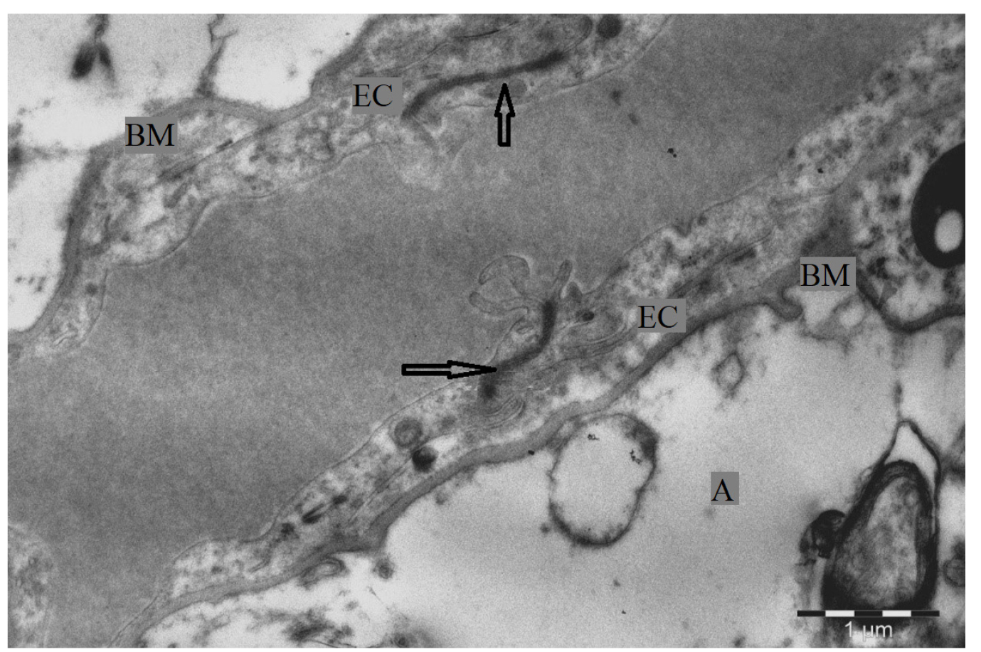
Figure 1. Ultrastructure of the basal membrane of the cortical capillary mammalian cerebellum. Electron micrograph: EC – endothelial cell; A – astrocyte; BM – basal membrane; → – tight junctions.
In some cells, the the nuclear surface has a tortuous shape due to the undulations of the karyolemma. (Figure 2) The nuclear envelope consists of two lipoprotein membranes, each 7 to 8 nm thick. The perinuclear space lies between the membranes. The membrane sometimes contains pores with diameters ranging from 70 to 100 nm. The central part of the matrix is usually electron light. Along the periphery of the nucleus, electron-dark accumulations of heterochromatin are noticeable, contacting the internal karyolemma.
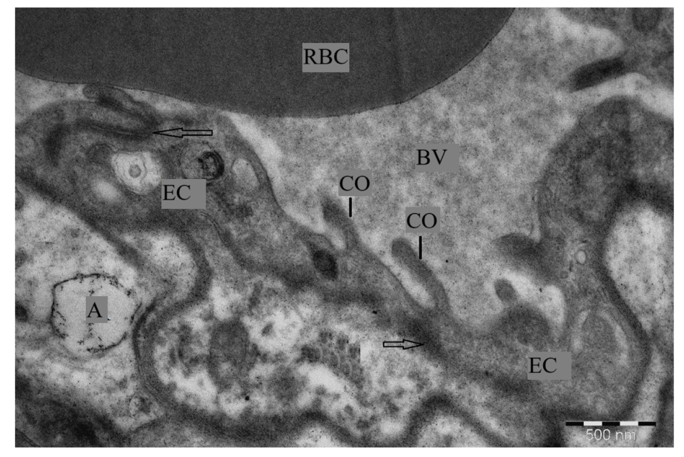
Figure 2. Ultrastructure of the blood-brain barrier of the capillary of the cerebellar cortex of a domestic bull. Electron micrograph: EC– endothelial cell; P– pericyte; A– astrocyte; BV– blood vessel lumen; E– erythrocyte; → – basal membrane.; PL– phagolysosomes; M– mitochondria.
The cytoplasm occupies the marginal part of the cell. Its thickness is slightly lower than the central part, the region occupied by nucleus, and varies within 1.3–1.8 μm. Electron-light mitochondria are detected in the cytoplasm. They contain thin cristae formed by folds of the inner membrane. The outer membrane of the organelles forms their shape, which varies from round to oval. The diameter of the mitochondria is about 1.0 μm. Also the cytoplasm contained a significant number of ribosomes, which look like electron-dense granules with a diameter of 25 to 30 nm. In addition to individual ribosomes, polyribosomes, ribosomes clusters, as well as a small number of pinocytotic vesicles and single short channels of the granular endoplasmic reticulum were identified (figure 3).
Figure 3. Ultrastructure of the endothelial cell of the capillary of the cerebellar cortex of a domestic bull. Electron micrograph: EC – endothelial cell; P– pericyte; A– astrocyte; BV– blood vessel lumen; GER – granular endoplasmic reticulum; → – basal membrane.
The cytoplasmic membrane of endothelial cells formed their outer shell, and its thickness varies within 7.5–10.0 nm in the obtained electronograms. In accordance with the location of the endotheliocytes, it forms their luminal and basal surfaces. Luminal surface facing the inside of the vessel. With an increase in the functional activity of the endothelial cell, it is able to form outgrowths and protrusions. The basal surface of the cells contacts the basal membrane (Figure 4).
Figure 4. Ultrastructure of a mammalian cerebellar cortex capillary endothelial cell in the active phase. Electron microphotography: EC—endothelial cell; A– astrocyte stalk; BV– blood vessel lumen; CO – endothelial cell cytoplasmic outgrowths; RBC– red blood cell; → – tight junctions.
Discussion
The present study revealed several key features and ultrastructural characteristics of endothelial cells that deserve detailed discussion.
First, the endothelial cells of the bovine cerebellar cortex capillaries exhibit a high mitochondrial density. This feature indicate an intensive level of metabolic activity, likely reflecting the high energy requirements of endothelial cells associated with transport of nutrients and oxygen to cerebellar neurons. [ 10 , 15 ] Similar mitochondrial abundance has been reported of other mammalian species, emphasizing the universality of this phenomenon. [ 16 - 20 ]
Second, the obtained data indicate the presence of numerous pinocytotic vesicles within the endothelial cytoplasm. This indicates active transport of substances through the endothelium, which plays a key role in maintaining homeostasis of the cerebellar microenvironment. The variability in the number and size of pinocytotic vesicles may be influenced by the physiological state of the animal. [ 9 , 11 , 18 ]
The third important aspect is the presence of tight junctions between endotheliocytes, which form the blood-brain barrier. These junctions play a critical role in preventing potentially harmful substances from entering the brain from the blood [ 8 , 17 ]. The results show that tight junctions in the capillaries of the bovine cerebellar cortex are highly organized, which may be due to the need for strict control over the composition of the cerebellar microenvironment.
In addition, the presence of pericytes, which are closely associated with endothelial cells, is observed. Pericytes play an important role in regulating blood flow and maintaining the capillaries structural integrity [ 17 ]. Their presence in the capillaries of the bovine cerebellar cortex supports their critical role in microcirculation dynamics in this part of the brain.
Thus, the results confirm that endothelial cells of the cerebellar cortex capillaries of the domestic bull are the primary element of the blood-brain barrier. Forming the lumen of the capillary, they contact each other with tight junctions and lie on a continuous basal membrane. The continuity of the latter and the presence of tight junctions block the possibility of spontaneous transport of macromolecular compounds from the bloodstream to the brain tissue.
CONCLUSION
In conclusion, this study highlights the complex ultrastructural organization of endothelial cells in the cerebellar cortical capillaries of the domestic bull. Revealed features, including high mitochondrial density, active pinocytosis, well-developed tight junctions, and the presence of pericytes, indicate the specialization and high functional activity of these cells.
Declaration of Generative AI and AI-assisted technologies in the writing process
AI-Assisted technologies were not applied at all stages of the work process.
Ethical Approval
The experiment was approved by the ethical committee Saint Petersburg State University of Veterinary Medicine.
Five fragments of the cerebellar cortex tissue from an adult domestic bull were used in the study. Each specimen measured no more than 2.0 mm3. The tissue samples were pre-fixed in a 2.0% glutaraldehyde solution prepared in a cacodylate buffer (pH 7.2–7.4) for 2 hours. Following primary fixation, fragments were rinsed in three changes of the same buffer. Post-fixation was performed in a 1.0% solution of osmium (VIII) oxide (diluted in cacodylate buffer, pH 7.2–7.4) for 1 hour. After post-fixation, the samples were dehydrated through a graded series of ethanol solutions, and subsequently treated with absolute acetone. The dehydrated tissue fragments were then embedded in Epon-812 resin, in accordance with the generally accepted methodology [ 12 ]. From each embedded block, three ultrathin sections were obtained using an ultramicrotome (LKB-III, Sweden). The sections were contrasted with 2.0% aqueous uranyl acetate solution and then with lead citrate solution. Imaging was performed using a Jem-1011 electron microscope (JEOL, Japan) at magnifications ranging from 2500× to 3000× [ 13 ]. All terminology used in this study follow the International Histological Nomenclature [ 14 ].
Authors' Contributions
P.A.V. planned the experiments, carried out the experiment, contributed to the interpretation of the results. V.S.P. contributed to sample preparation, contributed to the interpretation of the results, took the lead in writing the manuscript. All authors provided critical feedback and helped shape the research, analysis and manuscript.
Acknowledgements
The authors extend their sincere thanks to the members of Federal State Budgetary Educational Institution of Higher Education “Saint-Petersburg State University of Veterinary Medicine” for their help and support.
Conflict of interest
Authors declare that they have no conflict of interest. There were no ethical considerations to be considered in this research.
References
- Prusakov AV. Features of X-ray anatomy of the arterial vascular bed of the brain of a dachshund [Russian]. Scientific notes of the Kazan State Academy of Veterinary Medicine named after N.E. Bauman. 2014; 218(2): 215-220.
- Prusakov A, Zelenevskiy N, Shchipakin M, Bylinskaya D, Barteneva Y, Vasilyev D, et al. Blood supply to the brain of the long-tailed chinchilla (Chinchilla lanigera) [Russian]. Ippology and veterinary science. 2019; 2(32): 90-93.
- Yashin AV, Prusakov AV. Features of the state of the microcirculatory bed and membrane digestion in newborn calves with dyspepsia [Russian]. International Bulletin of Veterinary Medicine. 2021; 2: 155-160.DOI
- Drozdova LI. Histohematic barriers and their role in pathology [Russian]. Omsk Scientific Bulletin. 2004; S1-1 (26): 179-180.
- Akmalova GM, Ron GI, Musina LA, Chernysheva ND. Morphological features of the structure of elements of histohematic barriers in diseases of the oral mucosa [Russian]. In the collection: Proceedings of the thirteenth Siberian Congress: "Dentistry and Maxillofacial Surgery with International Participation"; All-Russian Symposium: "New Technologies in Dentistry". 2018: 20-23.
- Prusakov AV, Zelenevsky NV, Shchipakin MV, Bylinskaya DS, Vasiliev DV. Comparative ultrastructural organization of the blood-brain barrier of birds and mammals [Russian]. Morphology -2019 No. 6 p. 114.
- Prusakov AV, Zelenevsky NV. Histohematic barriers of animals. St. Petersburg: Cultural and Educational Partnership, 2022: 46.
- Karelina TV. Age-related features of synaptic activation of cerebellar Purkinje cells by the system of climbing and mossy fibers: specialty 03.03.01 "Physiology": dissertation for the degree of candidate of biological sciences. [Russian]. St. Petersburg, 2010: 125.
- Lobanov SA. Ultrastructure of the cerebellum during hypoxia. Evidence of morphological studies in medicine. [Russian] Proceedings of the International Scientific and Practical Conference. 2024: 59-63.
- Lobanov SA, Oberyukhtina EV, Cherepanov NS, Mansurova ZR. Some morphological features of the cerebellum. [Russian] Bulletin of scientific conferences. 2024; 12-2(112): 50-51.
- Mazloev AB. Microstructural features of the cerebellar cortex in newborns. [Russian] Morphology. 2019; 155(2): 182-183.
- Prusakova AV, Zelenevskiy NV, Prusakov AV, Yashin AV, Ponamarev VS. Ultra-structural organization of the gallbladder mucous membrane of Anglo-Nubian goat. Vet Res Forum. 2024; 15(3): 165-169.DOI
- Prusakova AV, Zelenevskiy NV, Prusakov AV, Yashin AV, Ponamarev VS. Organization of histo-hematic barriers of the liver in Anglo-Nubian goat. Online Journal of Animal and Feed Research. 2023;13(4): 242-245.DOI
- Prusakova AV, Zelenevskiy NV, Prusakov AV, Yashin AV, Ponamarev VS. Ultrastructural organization of liver hepatocytes of the Anglo-Nubian goat. Veterinarski Glasnik. 2023; 77(2): 176-187.DOI
- Hayat MA. Principles and Techniques of Electron Microscopy: Biological Applications, 4th ed. Cambridge University Press: Cambridge, UK. 2000: 543.
- Electron Microscopy Sciences. Preparation of Post-Staining Solutions [technical protocol]. Hatfield, PA: Electron Microscopy Sciences. Accessed October 21, 2025. https://www.emsdiasum.com/docs/technical/protocols/KN_PostStainSol.pdf
- Semchenko VV, Samuseva RP, Moiseeva MV. International histological nomenclature [Russian]. Omsk: Kolosova, 1999: 156.
- Barkhatova VP. Neurotransmitter organization and functional significance of the cerebellum. [Russian] Annals of Clinical and Experimental Neurology. 2010; 4(3): 44-49.DOI
- Akhmedov UB. Morphological changes in dynamics after leg amputation in the foliate region of the cerebellar vermis in dogs. Re-Health Journal. 2020; 1(5): 96-110.DOI
- Esakov SA. Morphofunctional organization of motor representations of facial muscles in the cortex and central nuclei of the cerebellum of a white mouse: specialty 03.03.01 "Physiology": dissertation for the degree of candidate of biological sciences. [Russian] Izhevsk, 2003: 124.
- Strizhikov VK, Ponomareva TA. Anatomical characteristics of the cerebellum in poultry. [Russian] Eurasia-2022: social and humanitarian space in the era of globalization and digitalization: Proceedings of the International Scientific Cultural and Educational Forum. 2022: 389–391.
- Karnyushko OA, Zimatkin SM. Disturbances in the morphogenesis of the cerebellar cortex of the offspring of rats with experimental cholestasis and their correction. [Russian] Bulletin of the National Academy of Sciences of Belarus. Series of Medical Sciences. 2015; 3: 95-101.
- Luzin VI, Churilin OA, Zolotarevskaya MV. Morphological features of the rat cerebellar nuclei (literature review). [Russian] Morphological almanac named after V. G. Koveshnikov. 2020; 18(3): 97-101.


